
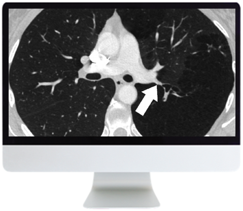
ARRS Basic Chest Imaging 2019

$30.00
Video Course · Pulmonary /Respiratory · 2019
The ARRS basic chest imaging course packs11 focused video lectures into two modules, covering atelectasis, pulmonary nodules, PE interpretation, and airways disease — core competencies for any radiologist reading chest studies.
-
11videos
-
2modules
-
2019edition
- Six expert lecturers cover the essential patterns in chest radiology
- Addresses atelectasis, infections in immunocompromised hosts, and pulmonary nodule management
- PE interpretation goes beyond filling defects to technique, pitfalls, and overdiagnosis
ARRS Basic Chest Imaging — Foundational Thoracic Radiology
The ARRS basic chest imaging programme delivers11 video lectures across two focused modules, distilling the core competencies every radiologist needs for chest study interpretation. From atelectasis patterns to pulmonary nodule management and PE pitfalls, this course builds the foundational vocabulary of thoracic radiology through six expert instructors from the American Roentgen Ray Society faculty.
-
11video lectures
-
2modules
-
2019edition
Inside this resource
ARRS Basic Chest Imaging: Fundamental Patterns
Module 1 opens with J. Czum’s systematic approach to atelectasis on chest radiographs and CT — distinguishing the patterns that matter clinically. R. Madan covers imaging of infections in immunocompromised hosts, a topic that demands both pattern recognition and clinical context. J. Ko then tackles pulmonary nodule management, addressing solid nodule characterization and the evolving concepts around adenocarcinoma and subsolid nodules.
Nodules, Airways, and Vascular Imaging
Module 2 shifts to broader pattern analysis. D. Manos reviews the nodular pattern on chest CT, building a differential framework for the radiologist. L. Haramati delivers a particularly practical session on pulmonary embolism — going beyond simple filling defects to cover technique, common pitfalls, misdiagnosis, and the growing concern of overdiagnosis. M. Hammer rounds out the course with imaging of airways disease.
Who this serves
- Radiology residents — building foundational chest imaging competency during training
- General radiologists — refreshing core thoracic patterns encountered in daily practice
- Clinicians involved in thoracic imaging — seeking structured education on chest CT and radiograph interpretation
Faculty & syllabus
Topics covered 11 sessions
- Module 1
- Spectrum of Atelectasis on Chest Radiographs and CT — J. Czum
- Imaging of Infections in Immunocompromised Hosts — R. Madan
- Management of Solid Pulmonary Nodules: Characterization and Update — J. Ko
- Adenocarcinoma and the Subsolid Nodule: Current Concepts — J. Ko
- Module 2
- Nodular Pattern on Chest CT — D. Manos
- Pulmonary Embolism: Beyond Filling Defects — L. Haramati
- Imaging of Airways — M. Hammer
Before you buy
Is this course suitable for experienced radiologists?
The content is designed as a foundational review. Experienced chest imagers may find value in the PE pitfalls and subsolid nodule sessions, which address nuances beyond basic pattern recognition.
The chart
| Format | 11 video lectures |
|---|---|
| Year | 2019 |
| Publisher | ARRS |
| Department | Pulmonary /Respiratory |
| Delivery | Instant download after checkout · lifetime re-access |
Verified original fileInstant downloadEncrypted checkout
Be the first to review “ARRS Basic Chest Imaging 2019”
Related products

Addiction Medicine
Oakstone Addiction Medicine for Non-Specialists 2025 (Videos + Slides PDFs)

Addiction Medicine
Providence Regional Medical Center Everett 28th Annual Fundamentals of Addiction Medicine 2026
Pulmonary /Respiratory
29th Annual Hypertension, Diabetes, & Dyslipidemia Conference 2025 (Videos + Slides)
Pulmonary /Respiratory
The Brigham Board Review and Comprehensive Update in Pulmonary Medicine 2022

Pulmonary /Respiratory
National Jewish Health 48th Annual Pulmonary & Allergy Update 2026
Pulmonary /Respiratory
The Brigham Board Review and Comprehensive Update in Pulmonary Medicine 2025 (Videos + Slides)
Reviews
There are no reviews yet.