
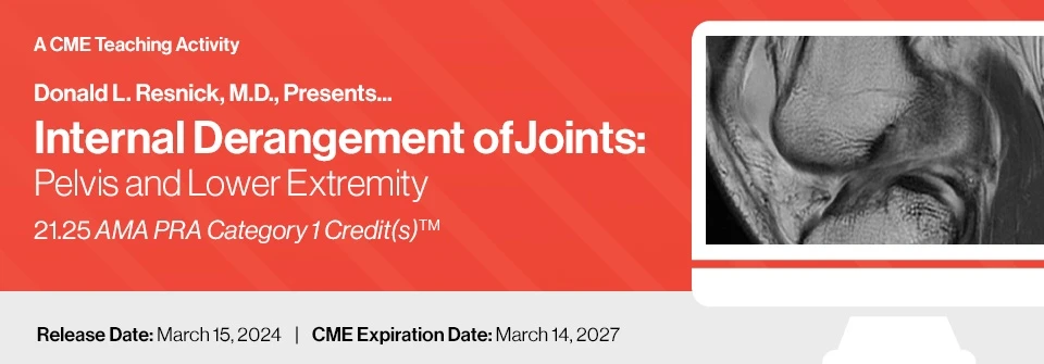
2024 Donald L. Resnick, M.D., Presents… Internal Derangement of Joints Pelvis and Lower Extremity

$50.00
VIDEO COURSE · Radiology · 2024
Dr. Donald Resnick presents 34 lectures on internal derangement joints pelvis and lower extremity — covering joint morphology, inflammatory and degenerative disorders, osteonecrosis versus insufficiency fractures, and tumor-like conditions with MR imaging correlation.
-
34lectures
-
1lead faculty
-
2024year
- MR imaging assessment of peripheral joint internal derangements
- Osteonecrosis versus insufficiency fractures of the hip and knee
- Inflammatory and degenerative joint disorders with imaging criteria
- Tumor and tumor-like conditions affecting bones and soft tissue
Donald Resnick on internal derangement of joints — pelvis and lower extremity
Dr. Donald Resnick’s 34-lecture series on internal derangement joints pelvis and lower extremity provides a systematic approach to MR imaging assessment of peripheral joint disorders. The course covers articular disorders of synovium-lined joints, focusing on joint morphology and general abnormalities, specific inflammatory and degenerative conditions, and tumor and tumor-like disorders — all with emphasis on the imaging criteria that guide clinical decision-making. Targeted at radiologists, orthopedic surgeons, rheumatologists, podiatrists, and sports medicine physicians.
-
34lectures
-
1lead faculty
-
2024year
Inside this resource
Joint morphology and articular disorders
The foundational sessions address joint morphology and general abnormalities seen across synovium-lined joints — establishing the anatomic framework that underpins accurate MR interpretation. Dr. Resnick walks through specific inflammatory and degenerative disorders of the hip, knee, and pelvis, connecting pathologic findings on MRI to the clinical questions that prompted the study. This structured approach builds the pattern recognition that radiologists and clinicians need for confident differential diagnosis.
Osteonecrosis versus insufficiency fractures
A dedicated focus session tackles one of the most common diagnostic dilemmas in MSK imaging: distinguishing osteonecrosis from insufficiency fractures, with particular emphasis on the hip and knee. Dr. Resnick presents the imaging features, distribution patterns, and clinical contexts that help separate these two entities — a distinction with direct implications for treatment planning and prognosis.
Tumors and tumor-like disorders
The tumor-focused sessions evaluate bone and soft tissue neoplasms, including entities that create diagnostic difficulty because of their imaging overlap with inflammatory or degenerative conditions. For each disease reviewed, the most current histologic and radiographic criteria are discussed, along with the role of molecular testing and immunohistochemistry when imaging alone is insufficient for definitive diagnosis.
Who this serves
- Radiologists — comprehensive MR imaging criteria for pelvis and lower extremity joint disorders
- Orthopedic surgeons — imaging-based decision support for surgical planning in hip, knee, and pelvic conditions
- Rheumatologists — imaging correlation for inflammatory and degenerative joint disease assessment
The chart
| Format | 34 video lectures |
|---|---|
| Faculty | Donald L. Resnick, MD |
| Scientific Sponsor | Educational Symposia |
| Publisher | Educational Symposia |
| Department | Orthopedics |
| Delivery | Instant download after checkout · lifetime re-access |
Verified original fileInstant downloadEncrypted checkout
5 reviews for 2024 Donald L. Resnick, M.D., Presents… Internal Derangement of Joints Pelvis and Lower Extremity
Add a review
Related products



Orthopedics

Thomas W. –
Dr. Resnick’s lectures are the gold standard for MSK imaging education. The osteonecrosis versus insufficiency fracture focus session alone is worth the price — it clarified distinctions I had been struggling with for years.
Michael C. –
This is the definitive resource for understanding internal derangement on MRI. Dr. Resnick’s systematic approach to articular disorders has changed how I read MSK imaging studies. Essential for any radiologist.
Sandra L. –
Comprehensive coverage of pelvis and lower extremity joint pathology. The joint morphology sessions provide an excellent foundation before moving into the disease-specific lectures. Would have liked more case material in the tumor section.
David K. –
Dr. Resnick connects imaging findings to clinical management in a way that few educators can. The hip and knee focus sessions are indispensable for anyone interpreting MSK MRI. Highly recommended.
Jennifer H. –
Outstanding lectures from a legendary educator in MSK radiology. The inflammatory and degenerative disorder sections are particularly well-organized. The 34 lectures cover a lot of ground but remain digestible.