DentistryOnline courseGnathology
Reading dental images with confidence — radiographic anatomy, panoramic errors, CBCT, jaw cysts, tumours and TMJ imaging
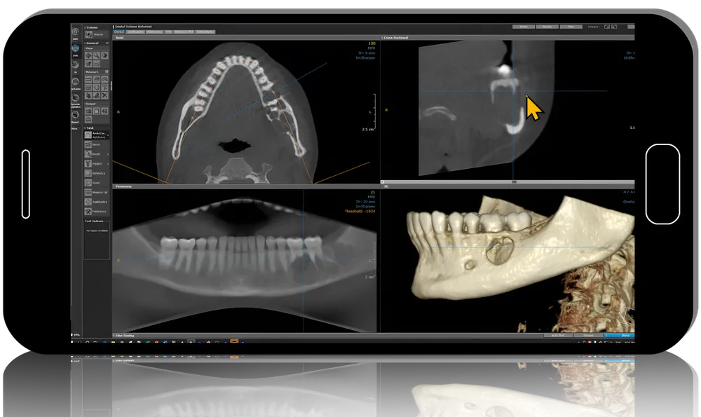
Oral and maxillofacial radiologist Mansour Ahmad, trained at the University of Connecticut Health Center, covers Oral and Maxillofacial Radiology: Basics to Advanced in 16 lessons lasting 7 h 25 min. The course moves from interpretation principles and normal intraoral, panoramic and CBCT anatomy to positioning errors, cysts, benign tumours, soft tissue calcifications and imaging of the temporomandibular joint, with five short panoramic case readings.
- 16lessons
- 7 h 25 minof video
- 5panoramic case readings
What you’ll be able to do
- Apply a systematic approach to radiographic interpretation
- Identify normal landmarks on intraoral, panoramic and CBCT images
- Spot and correct common panoramic positioning errors
- Differentiate jaw cysts and benign tumours by their radiographic features
- Recognise soft tissue calcifications and choose imaging for the TMJ
Inside the course
Normal anatomy first
Lessons on intraoral anatomy of each jaw, panoramic anatomy and basic CBCT anatomy build the reference picture needed before judging anything abnormal.
Image quality and interpretation
A 39-minute lesson on panoramic positioning faults sits beside the principles of interpretation, followed by five brief real-case panoramic readings.
Pathology and the joint
Two parts on cysts of the jaws (about 100 minutes), an hour on benign tumours, 40 minutes on calcifications and 52 minutes on TMJ imaging form the advanced half.
Built for
- General dentistsSharpens everyday reading of periapicals and panoramics.
- Dental students and residentsA structured review of radiology topics from anatomy to lesions.
- Clinicians starting with CBCTAn anatomical orientation before interpreting 3D scans.
Curriculum
Course lessons (7 h 25 min) 16 lessons
- Cyst of Jaw Part 1 (51 min)
- Cyst of Jaw Part 2 (49 min)
- Panoramic Radiographic Interpretation Case 1 (9 min)
- Panoramic Radiographic Interpretation Case 2 (6 min)
- Panoramic Radiographic Interpretation Case 3 (6 min)
- Panoramic Radiographic Interpretation Case 4 (8 min)
- Panoramic Radiographic Interpretation Case 5 (9 min)
- Basic CBCT (Cone Beam CT) Anatomy (20 min)
- Benign Tumors (1 h 4 min)
- Imaging of the TMJ (52 min)
- Intraoral Radiographic Anatomy of the Mandible (21 min)
- Intraoral Radiographic Anatomy of the Maxilla (25 min)
- Panoramic Radiographic Anatomy (17 min)
- Positioning Errors in Panoramic Radiography (39 min)
- Principles of Radiographic Interpretation (29 min)
- Soft Tissue Calcifications of the Maxillofacial Region (40 min)
Before you buy
In what order should I watch the lessons?
The files are listed with pathology first; starting with the principles and anatomy lessons, then moving to cases and lesions, gives a more logical path.
Does it cover malignant tumours or implant planning on CBCT?
Neither appears in the lesson list; pathology is limited to cysts, benign tumours and calcifications.
Do I get CME credit or a certificate?
No. Clinicapedia provides the educational content only — no CME or CE credits, MOC points or certificates of completion are included with this product.
Specifications
| Format | Online video course |
|---|---|
| Instructor | Mansour Ahmad |
| Contents | 16 lessons |
| Duration | 7 h 25 min |
| Access | Lifetime, self-paced |
| Department | Dentistry |
| Delivery | Instant download · lifetime re-download |
Related products




Anesthesiology