
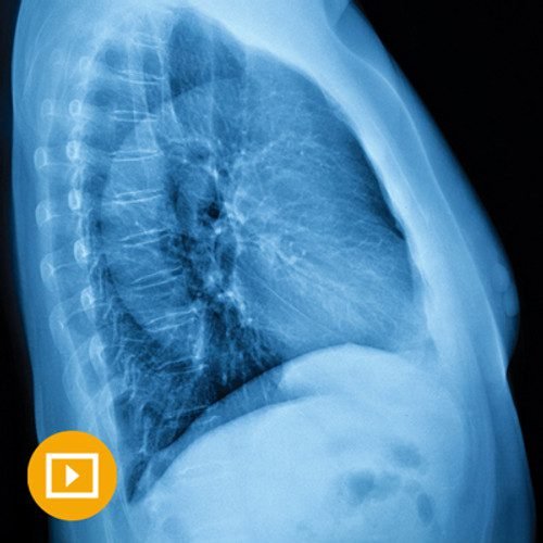
UCSF Thoracic Imaging 2023

$35.00
Video Course · Pulmonary /Respiratory · 2023
The UCSF thoracic imaging course provides30 focused lectures on CT, PET, and MRI interpretation across the pulmonary, vascular, and cardiac systems — delivered by four UCSF radiology faculty.
-
30lectures
-
12.25hours
-
4speakers
-
2023edition
- Practical approach to high-resolution CT, PET-CT, and MRI of the chest
- Covers lung cancer screening, pulmonary nodules, and updated interstitial pneumonia criteria
- Addresses acute vascular emergencies including PE and aortic syndromes
UCSF Thoracic Imaging Course 2023 — Practical CT, PET, and MRI Review
The UCSF thoracic imaging course is a 12.25-hour programme that delivers30 lectures covering the full spectrum of chest imaging. Led by Brett M. Elicker, Maya Vella, Kimberly G. Kallianos, and Brian M. Haas, the curriculum emphasizes practical CT interpretation — including PET and MRI correlations — for radiologists who read thoracic studies in daily practice.
-
30video lectures
-
12.25hours
-
4UCSF faculty
-
2023edition
Inside this resource
UCSF Thoracic Imaging Course: Diffuse Lung Disease and Chest Radiographs
The opening section tackles the hardest patterns in chest imaging — fibrotic lung disease, mosaic attenuation, cystic disease, and chronic consolidation. Elicker and Vella walk through a multidisciplinary approach to diffuse lung disease, while Kallianos and Haas cover challenging chest radiographs, lateral film analysis, and post-operative findings. Challenging case conferences bookend each module.
Neoplasms, Nodules, and Infection
Lung nodule management receives dedicated attention with updates on subsolid nodules, incidentalomas, and CT-guided biopsy technique. Elicker leads the neoplasm and infection sessions, covering pulmonary nodule approach, mediastinal masses, and classic appearances of unusual infections. Haas unpacks the tuberculosis chest radiograph and mediastinal contour analysis.
Vascular and Cardiac Diseases
Kallianos anchors the vascular section with updates on PE interpretation, acute aortic syndromes, and CT findings in pulmonary hypertension. Vella covers beyond-PE acute chest pain findings, cardiothoracic hardware, and post-operative cardiac assessment. Congenital heart disease repair evaluation and thoracic vascular trauma complete the cardiovascular curriculum.
Who this serves
- Radiologists — interpreting thoracic imaging studies who need a structured, practical update
- Radiology residents and fellows — building foundational and advanced chest imaging competency
- Pulmonologists — reviewing imaging patterns and updated diagnostic criteria for lung diseases
Faculty & syllabus
Faculty 4 speakers
- Brett M. Elicker, MD — diffuse lung disease, lung nodules, infections, mediastinal masses, CT-guided biopsies
- Maya Vella, MD — fibrotic lung disease, airways disease, acute chest pain, cardiothoracic hardware
- Kimberly G. Kallianos, MD — cystic lung disease, PE, aortic syndromes, pulmonary hypertension, congenital heart disease
- Brian M. Haas, MD — chronic consolidation, chest radiograph analysis, thoracic trauma, tuberculosis imaging
Topics covered 30 lectures
- Multi-disciplinary Approach to Diffuse Lung Disease
- An Approach to Distinguishing Fibrotic Lung Diseases
- Cystic Lung Disease
- Mosaic Attenuation-Perfusion
- Airways Diseases
- Chronic Consolidation
- Challenging Chest Radiographs
- Iatrogenesis Imperfecta — Post-Op Findings in the Chest
- Elevating the Analysis of the Lateral Chest Radiograph
- Challenging Chest Cases (Diffuse Lung Disease)
- Update on the Approach to the Pulmonary Nodule
- Thoracic Incidentalomas
- The Subsolid Nodule — Current Concepts
- Mediastinal Contours on the Chest Radiograph
- CT-Guided Lung Biopsies — Approach, Risks, and Benefits
- Approach to Pulmonary Infections
- Classic Appearances of Unusual Infections
- Unpacking the Tuberculosis Chest Radiograph
- Approach to the Mediastinal Mass
- Challenging Chest Cases (Neoplasms)
- Update on Pulmonary Embolism
- Beyond No PE — CT Findings of Acute Chest Pain
- Thoracic Vascular Trauma
- Acute Aortic Syndromes
- Cardiothoracic Hardware Challenge
- Approach to Repaired Congenital Heart Disease
- Post-op Heart — Normal Findings and When to Raise the Alarm
- CT Clues in Pulmonary Hypertension
- Congenital Lung Lesions
- Challenging Chest Cases (Vascular)
Before you buy
Who is the intended audience?
Radiologists, physician assistants, and nurses involved in thoracic imaging interpretation. The content ranges from practical pattern recognition to advanced diagnostic reasoning.
What imaging modalities are covered?
The course focuses on CT including PET-CT, with MRI and radiography correlations throughout. High-resolution CT technique is a recurring emphasis across all three sections.
The chart
| Format | Video lectures |
|---|---|
| Duration | 12.25 hours |
| Lectures | 30 |
| Year | 2023 |
| Publisher | UCSF |
| Department | Pulmonary /Respiratory |
| Delivery | Instant download after checkout · lifetime re-access |
Verified original fileInstant downloadEncrypted checkout
Be the first to review “UCSF Thoracic Imaging 2023”
Related products

Pulmonary /Respiratory
National Jewish Health 48th Annual Pulmonary & Allergy Update 2026

Addiction Medicine
Harvard Principles of Medical Education: Maximizing Your Teaching Skills 2024 -October Edition
Pulmonary /Respiratory
OnExamination SCE In Respiratory Medicine (Updated Feb 2023) (PDF)

Pulmonary /Respiratory
29th Annual Hypertension, Diabetes, & Dyslipidemia Conference 2025 (Videos + Slides)
Reviews
There are no reviews yet.