
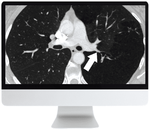
ARRS Basic Chest Imaging CME Course 2019

$35.00
Online Course · Radiology · 2019
The ARRS Basic Chest Imaging CME Course 2019 reviews 11 lectures (1.9GB) on atelectasis, pulmonary nodule management, pulmonary embolism, and airway imaging — designed for radiologists updating their thoracic imaging skills.
Chest Imaging CME Course Covering Nodules, Embolism, and Airway Disease
The ARRS Basic Chest Imaging CME Course 2019 is an ARRS online course of 11 lectures (1.9GB) offering a focused review of basic chest imaging topics and updates in thoracic disease. Coverage spans imaging of airways, atelectasis, infections in immunocompromised hosts, pulmonary nodule management, and pulmonary embolism.
-
11Lectures
-
2Modules
-
2019Edition
Inside this resource
Atelectasis & Immunocompromised Infections
J. Czum reviews the spectrum of atelectasis on chest radiographs and CT, while R. Madan covers imaging of infections in immunocompromised hosts.
Pulmonary Nodule Management
J. Ko presents current concepts and updates on management of solid pulmonary nodules along with adenocarcinoma and the subsolid nodule.
Nodular Patterns & Pulmonary Embolism
D. Manos discusses nodular patterns on chest CT and L. Haramati covers pulmonary embolism diagnosis, pitfalls, misdiagnosis, and overdiagnosis.
Airway Imaging
M. Hammer closes the course with imaging of airways, including protocols, tracheal and bronchial tumors, tracheomalacia, and bronchiectasis.
Who this serves
- Radiologists reviewing basic chest imaging topics — the course is framed as a focused review of basic chest imaging topics and thoracic disease updates.
- Radiologists managing pulmonary nodules and embolism cases — modules address current guidelines for nodule management and pitfalls in pulmonary embolism diagnosis.
Faculty & syllabus
Faculty 6 speakers
Lectures are delivered by named thoracic imaging faculty across two modules.
- J. Czum — Spectrum of atelectasis on chest radiographs and CT.
- R. Madan — Imaging of infections in immunocompromised hosts.
- J. Ko — Management of solid pulmonary nodules and the subsolid nodule/adenocarcinoma spectrum.
- D. Manos — Nodular pattern on chest CT.
- L. Haramati — Pulmonary embolism technique, pitfalls, misdiagnosis, and overdiagnosis.
- M. Hammer — Imaging of airways, including tracheal/bronchial tumors and tracheomalacia/bronchiectasis.
Before you buy
What does the ARRS Basic Chest Imaging CME course cover?
It covers 11 lectures (1.9GB) across 2 modules addressing atelectasis, immunocompromised infections, pulmonary nodule management, nodular CT patterns, pulmonary embolism, and airway imaging.
Who are the course faculty?
Named faculty include J. Czum, R. Madan, J. Ko, D. Manos, L. Haramati, and M. Hammer, each covering a specific thoracic imaging topic.
The chart
| Format | Online course (video lectures) |
|---|---|
| Duration | 11 files · 1.9GB |
| Faculty | 6 named speakers |
| Year | 2019 |
| Publisher | ARRS |
| Department | Radiology |
| Delivery | Instant download after checkout · lifetime re-access |
Verified original fileInstant downloadEncrypted checkout
Be the first to review “ARRS Basic Chest Imaging CME Course 2019”
Related products


Radiology


Radiology
Radiology
Reviews
There are no reviews yet.