
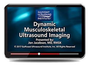
Gulfcoast Dynamic Musculoskeletal Ultrasound Imaging (Videos+PDFs)
$15.00
VIDEO COURSE + PDFS · Gulfcoast Ultrasound Institute
This dynamic MSK ultrasound imaging course from Gulfcoast demonstrates real-time pathology across every major joint — from shoulder biceps subluxation and snapping hip syndrome through Achilles tendon pathology and peroneal dislocation. Thirty-four conditions covered with dynamic maneuver demonstrations.
-
1training video
-
34pathology stations
-
7joint regions
- Shoulder: biceps subluxation, impingement, adhesive capsulitis
- Elbow: ulnar nerve dislocation, snapping triceps, UCL tears
- Knee: meniscal tears, patellar clunk, snapping tendons
- Ankle/foot: peroneal dislocation, Achilles pathology, Morton neuroma
Dynamic MSK ultrasound imaging for joint and soft tissue pathology
This dynamic MSK ultrasound imaging course from Gulfcoast is a comprehensive demonstration of musculoskeletal pathology evaluated with real-time dynamic maneuvers. Thirty-four conditions across seven body regions are presented — shoulder, elbow, wrist and hand, hip and thigh, knee, ankle and foot, and soft tissues. Each station demonstrates the pathology under dynamic loading or movement, showing findings that static imaging alone cannot capture.
-
34pathology stations
-
7joint regions
Inside this resource
Shoulder, elbow, and wrist dynamic assessment
The upper extremity modules demonstrate biceps brachii tendon subluxation and dislocation, impingement syndrome, and adhesive capsulitis with dynamic maneuvers. Elbow coverage addresses ulnar nerve dislocation, snapping triceps syndrome, and ulnar collateral ligament injuries from partial to complete tears. Wrist and hand pathology includes extensor carpi ulnaris dislocation, boxer knuckle, pulley tears, trigger finger, and gamekeeper’s thumb.
Hip, knee, and ankle/foot pathologies
Lower extremity stations cover snapping hip syndrome of the iliopsoas and iliotibial tract, patellar tendon tears, meniscal displacement and bucket handle tears, patellar clunk syndrome, and snapping semitendinosus and sartorius tendons. Ankle and foot coverage addresses peroneal tendon dislocation, tendon impingement, muscle hernia, Achilles tendon pathology, and Morton neuroma.
Soft tissue and special applications
A final station covers slipping rib syndrome — a soft tissue condition that requires dynamic assessment for accurate identification. The course includes supplementary PDF materials for reference alongside the video demonstrations.
Who this serves
- Sonographers — master dynamic scanning techniques that reveal pathology missed on static images
- Orthopedists and sports medicine physicians — learn to recognize dynamic findings for pre-surgical planning
- PM&R and rheumatology clinicians — add dynamic MSK assessment to the point-of-care ultrasound toolkit
Faculty & syllabus
Pathology stations 34 conditions
- Biceps Brachii Tendon Subluxation/Dislocation
- Impingement Syndrome
- Adhesive Capsulitis
- Ulnar Nerve Dislocation
- Snapping Triceps Syndrome
- Ulnar Collateral Ligament Partial Tear, Ulnar Collateral Ligament Laxity
- Ulnar Collateral Ligament Complete Tear
- Extensor Carpi Ulnaris Dislocation
- Boxer Knuckle
- Pulley Tear
- Trigger Finger
- Ganglion Cyst
- Gamekeeper’s Thumb
- Snapping Hip Syndrome (Iliopsoas; Iliotibial Tract)
- Patellar Tendon Full Thickness Tear
- Meniscal Displacement
- Bucket Handle Tear Medial Meniscus
- Intra-Articular Body
- Patellar Clunk Syndrome
- Snapping Semitendinosus Tendon
- Snapping Sartorius
- Peroneal Tendon Dislocation/Subluxation
- Tendon Impingement
- Muscle Hernia
- Achilles Tendon
- Morton Neuroma
- Slipping Rib Syndrome
Before you buy
Does this include PDFs?
Yes — supplementary PDF materials ship alongside the training video for reference.
How are the pathologies organized?
Thirty-four conditions are grouped by joint region — shoulder, elbow, wrist/hand, hip/thigh, knee, ankle/foot, and soft tissues.
The chart
| Format | 1 training video (MP4) + PDFs |
|---|---|
| Conditions | 34 pathology stations |
| Publisher | Gulfcoast Ultrasound Institute |
| Department | Gulfcoast Ultrasound Institute |
| Delivery | Instant download after checkout · lifetime re-access |
Verified original fileInstant downloadEncrypted checkout
5 reviews for Gulfcoast Dynamic Musculoskeletal Ultrasound Imaging (Videos+PDFs)
Add a review
Related products

Gulfcoast Ultrasound Institute
Gulfcoast Ultrasound Institute
GCUS Introduction to Peripheral Vascular Ultrasound 2022 (VIDEOS)

Gulfcoast Ultrasound Institute
Gulfcoast Ultrasound Institute
Advanced Musculoskeletal (MSK) Ultrasound Applications 2020 (VIDEOS)
Gulfcoast Ultrasound Institute
Gulfcoast Ultrasound Institute
GCUS Abdominal and Primary Care Ultrasound 2022 (Gulfcoast Ultrasound Institute) (Videos) 2022
Gulfcoast Ultrasound Institute
AIUM Prenatal Ultrasound Abnormalities of the Chest, Abdomen, and Pelvis With Postnatal Correlation
Gulfcoast Ultrasound Institute
Gulfcoast Upper Extremity Musculoskeletal Ultrasound Protocols (Videos)
Aisha B. –
Good variety of conditions with real-time maneuvers. The wrist and hand section covers pathology I encounter regularly. PDFs are a nice bonus.
M. Alvarez –
Comprehensive dynamic MSK course. The shoulder and knee sections are strongest. The UCL partial versus complete tear comparison was particularly instructive.
Sarah K. –
Thirty-four pathology stations across seven regions is an incredible resource. The snapping hip and patellar clunk demonstrations are outstanding for dynamic assessment learning.
Marcus J. –
Extensive pathology list but some stations move quickly through the dynamic maneuvers. Going back to review them at slower speed was key. The knee module is the strongest section.
drtomh –
Downloaded for the ankle and foot stations. The peroneal tendon dislocation and Achilles pathology demonstrations show exactly what dynamic scanning reveals.