
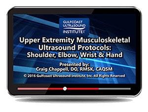
Gulfcoast Upper Extremity Musculoskeletal Ultrasound Protocols (Videos)


$15.00
VIDEO COURSE · Gulfcoast Ultrasound Institute
This upper extremity MSK ultrasound training video provides live demonstrations of standard protocols for evaluating the shoulder, elbow, hand, and wrist. A picture-in-picture format lets you watch the transducer position and ultrasound image simultaneously.
-
1training video
-
55 minruntime
-
20protocol stations
- Shoulder exam: rotator cuff tendons, biceps, AC and glenohumeral joints
- Elbow exam: anterior and posterior views, ulnar nerve assessment
- Wrist and hand: median nerve evaluation
- Picture-in-picture format for simultaneous transducer and image viewing
Upper extremity MSK ultrasound protocols for shoulder, elbow, and wrist
This upper extremity MSK ultrasound training video from Gulfcoast delivers live demonstrations of the standard scanning protocols used to evaluate the shoulder, elbow, hand, and wrist. Each station is presented in a picture-in-picture format — the participant sees the transducer position on the patient alongside the real-time ultrasound image, making the technique directly transferable to clinical scanning.
-
1training video
-
55 minruntime
-
20protocol stations
Inside this resource
Shoulder examination protocols
The shoulder module covers normal ultrasound anatomy across the biceps tendon, subscapularis, supraspinatus, infraspinatus, rotator cuff interval, acromioclavicular joint, and glenohumeral joint. Normal ultrasound characteristics are demonstrated alongside the shoulder impingement syndrome evaluation.
Elbow, wrist, and hand scanning
Elbow protocols address anterior and posterior views in both short and long axis, medial epicondyle assessment, and ulnar nerve evaluation including subluxation testing. The wrist and hand module covers median nerve scanning — completing the upper extremity protocol set.
Who this serves
- Sonographers — learn standardized upper extremity MSK protocols with real-time technique demonstrations
- Radiologists and orthopedists — review normal ultrasound anatomy and scanning positions for shoulder, elbow, and wrist
- Sports medicine and PM&R physicians — develop MSK ultrasound scanning skills for point-of-care evaluation
Faculty & syllabus
Protocol stations 20 stations
- Normal Ultrasound Anatomy and Protocols for the Shoulder, Elbow, Hand/Wrist
- Normal Ultrasound Characteristics for the Shoulder, Elbow, Hand/Wrist
- Biceps Tendon
- Subscapularis Tendon
- Rotator Cuff
- Supraspinatus Tendon
- Rotator Cuff Interval
- Infraspinatus Tendon
- Acromioclavicular Joint
- Glenohumeral Joint
- Shoulder Impingement Syndrome
- Anterior Elbow Views (Short and Long Axis)
- Medial Epicondyle
- Posterior Elbow Views (Short and Long Axis)
- Ulnar Nerve
- Ulnar Nerve Subluxation
- Median Nerve
Before you buy
What is the picture-in-picture format?
Each protocol station shows the transducer position on the patient and the corresponding ultrasound image side by side in real time.
What is the runtime?
One training video at 55 minutes covering20 protocol stations across the shoulder, elbow, hand, and wrist.
The chart
| Format | 1 training video (MP4) |
|---|---|
| Runtime | 55 minutes |
| Publisher | Gulfcoast Ultrasound Institute |
| Department | Gulfcoast Ultrasound Institute |
| Delivery | Instant download after checkout · lifetime re-access |
Verified original fileInstant downloadEncrypted checkout
5 reviews for Gulfcoast Upper Extremity Musculoskeletal Ultrasound Protocols (Videos)
Add a review
Related products

Gulfcoast Ultrasound Institute
AIUM VIDEO HD Musculoskeletal Ultrasound of Sports Injuries and Hernias

Gulfcoast Ultrasound Institute

Gulfcoast Ultrasound Institute
Gulfcoast Ultrasound Institute
GCUS Introduction to Peripheral Vascular Ultrasound 2022 (VIDEOS)
Gulfcoast Ultrasound Institute
Gulfcoast Ultrasound Institute
Gulfcoast Ultrasound Institute
GCUS Introduction to Transcranial Doppler Ultrasound 2019 (VIDEOS)

Gulfcoast Ultrasound Institute
Fatima R. –
Content is solid but the wrist and hand section is brief compared to the shoulder coverage. The median nerve station could use more detail.
Cathy N. –
Good coverage of shoulder and elbow protocols. The ulnar nerve subluxation evaluation was new to me. The55-minute runtime is manageable.
Raj P. –
Exactly what I needed for upper extremity scanning. The rotator cuff stations are well-organized and the medial epicondyle assessment is thorough.
Daniel K. –
The picture-in-picture format is outstanding for learning technique. Watching the transducer position alongside the ultrasound image makes the shoulder protocols click.
Jason Y. –
Downloaded for the elbow protocols. The anterior and posterior view demonstrations in both short and long axis are well-presented. Good reference material.