
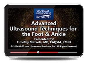
Gulfcoast Advanced Ultrasound Techniques for the Foot and Ankle (Videos+PDFs)
$15.00
VIDEO COURSE + PDFS · Gulfcoast Ultrasound Institute
This foot ankle ultrasound techniques course from Gulfcoast provides an advanced regional scan approach organized by anatomical compartment — lateral, posterolateral, anterolateral, and posterior — covering ligaments, peroneal tendons, the Achilles, and specialized injection techniques.
-
1training video
-
4anatomical compartments
-
24scan stations
- Lateral compartment: calcaneofibular ligament, subtalar joint, peroneal tendons
- Posterolateral: peroneals, superior retinaculum, low-lying brevis
- Anterolateral: ATFL, AITFL, gutter evaluation, Walin lesion
- Posterior: Achilles, Kager’s fat pad, retrocalcaneal bursa, os trigonum injections
Foot ankle ultrasound techniques for advanced regional scanning
This foot ankle ultrasound techniques course from Gulfcoast provides an advanced regional approach to foot and ankle ultrasound, organized by anatomical compartment. Twenty-four scan stations cover the lateral, posterolateral, anterolateral, and posterior compartments — from ligament integrity and peroneal tendon assessment through specialized injection techniques. Each station addresses both static and dynamic evaluation where applicable.
-
24scan stations
-
4anatomical compartments
Inside this resource
Lateral and posterolateral compartment scanning
The lateral module covers the calcaneofibular ligament with static and dynamic assessment, subtalar joint evaluation, inframalleolar and distal peroneal tendons, and the os peroneum. Posterolateral stations address the peroneal tendons, superior peroneal retinaculum, low-lying peroneus brevis, and quartus muscle belly identification.
Anterolateral and posterior compartment evaluation
Anterolateral scanning covers the anterior talofibular ligament and anterior inferior tibiofibular ligament in both static and dynamic views, the anterolateral gutter, and soft tissue or bony Walin lesions. The posterior module addresses the Achilles tendon, Kager’s fat pad, retrocalcaneal bursa impingement, os trigonum injection technique, and subtalar joint assessment.
Who this serves
- Sonographers — develop advanced foot and ankle scanning competency organized by anatomical compartment
- Orthopedists and podiatrists — review ligament and tendon assessment for ankle sprain and chronic pain evaluation
- Sports medicine physicians — add specialized foot and ankle ultrasound to the MSK diagnostic toolkit
Faculty & syllabus
Scan stations 24 stations
- Calcaneofibular Ligament (Static and Dynamic)
- Subtalar Joint
- Inframalleolar Peroneal
- Distal Peroneals
- Osperoneum
- Peroneals
- Superior Peroneal Retinaculum
- Low Lying Brevis
- Quartus Muscle Belly
- Anterior Talofibular Ligament (Static and Dynamic)
- AITFL (Static and Dynamic)
- Anterolateral Gutter
- Soft Tissue or Bony Walin Lesion
- Achilles
- Kager’s Fat Pad
- Retrocalcaneal Bursa Impingement
- Os Trigom Injections
- Subtalar Joint
Before you buy
Does this cover injection techniques?
Yes — the posterior module includes os trigonum injection technique with ultrasound guidance.
Does this include PDFs?
Yes — supplementary PDF materials are included alongside the training video.
The chart
| Format | 1 training video (MP4) + PDFs |
|---|---|
| Stations | 24 scan stations across 4 compartments |
| Publisher | Gulfcoast Ultrasound Institute |
| Department | Gulfcoast Ultrasound Institute |
| Delivery | Instant download after checkout · lifetime re-access |
Verified original fileInstant downloadEncrypted checkout
5 reviews for Gulfcoast Advanced Ultrasound Techniques for the Foot and Ankle (Videos+PDFs)
Add a review
Related products
Gulfcoast Ultrasound Institute
AIUM VIDEO HD Musculoskeletal Ultrasound of Sports Injuries and Hernias
Gulfcoast Ultrasound Institute

Gulfcoast Ultrasound Institute
Gulfcoast Ultrasound Institute
Gulfcoast Ultrasound Guided Prolotherapy: General Principles (Videos)
Gulfcoast Ultrasound Institute
AIUM Prenatal Ultrasound Abnormalities of the Chest, Abdomen, and Pelvis With Postnatal Correlation
Gulfcoast Ultrasound Institute

Gulfcoast Ultrasound Institute

Gulfcoast Ultrasound Institute
Wei L. –
Comprehensive content but some stations could use more pathology examples alongside the normal anatomy. The Walin lesion coverage is a unique addition.
M. Alvarez –
Advanced foot and ankle coverage that goes beyond basic protocols. The peroneal tendon and superior retinaculum stations filled gaps in my scanning knowledge.
Sarah K. –
Organized by anatomical compartment which makes it easy to follow along during scanning. The ATFL static and dynamic assessment section is particularly well-demonstrated.
Olivia P. –
Good advanced resource for foot and ankle scanning. The retrocalcaneal bursa and Kager’s fat pad modules are well-presented. PDFs supplement the video nicely.
Carlos M. –
The posterolateral compartment section on low-lying brevis and quartus muscle belly was exactly what I needed for chronic ankle pain evaluation.