
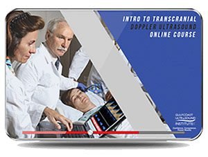
GCUS Introduction to Transcranial Doppler Ultrasound 2019 (VIDEOS)
$30.00
VIDEO COURSE · Gulfcoast Ultrasound Institute · 2019
This transcranial doppler ultrasound training program provides a comprehensive introduction to TCD and TCI examinations — from neuroanatomy and Doppler waveform characteristics through vasospasm, emboli monitoring, and patent foramen ovale evaluation.
-
6video lectures
-
3.00 GBlibrary size
-
21topics covered
-
2019edition
- TCD neuroanatomy with normal flow velocity ranges
- Doppler waveform analysis for vasospasm and subarachnoid hemorrhage
- Emboli monitoring and cerebral circulatory arrest detection
- PFO evaluation and sickle cell disease assessment
Transcranial doppler ultrasound training for TCD and TCI examinations
This transcranial doppler ultrasound training program from Gulfcoast provides a comprehensive review of how to perform and interpret TCD and TCI examinations. The curriculum covers neuroanatomy, Doppler waveform analysis, scan techniques, and diagnostic criteria aligned with AIUM and IAC guidelines. Twenty-one topics span from fundamental Doppler physics through clinical applications including vasospasm, emboli monitoring, sickle cell assessment, and patent foramen ovale detection.
-
6video lectures
-
3.00 GBlibrary size
-
21topics covered
-
2019edition
Inside this resource
Doppler physics and instrumentation fundamentals
The program opens with Doppler principles and the Doppler equation, interrogation angles, and the full range of instrumentation — pulsed wave, continuous wave, color Doppler, and power Doppler. Optimization of system controls and artifact recognition form the technical foundation before moving into neuroanatomy.
TCD neuroanatomy and waveform analysis
Normal intracranial flow characteristics and velocity ranges are covered alongside TCD window anatomy. The course addresses vasospasm evaluation, subarachnoid hemorrhage assessment, aneurysm rupture findings, and arteriovenous malformation identification — each paired with spectral waveform examples.
Clinical applications and advanced topics
Advanced sessions cover emboli monitoring, cerebral circulatory arrest, sickle cell disease assessment, traumatic head injury evaluation, and patent foramen ovale detection. TCD examination techniques for stenosis, collateral competency, and neurointervention round out the clinical curriculum.
Who this serves
- Sonographers — develop competency in TCD scanning techniques and waveform interpretation
- Neurologists — strengthen skills for evaluating vasospasm, emboli, and cerebrovascular disease
- Vascular surgeons and radiologists — add TCD/TCI proficiency to their diagnostic repertoire
Faculty & syllabus
Topics covered 21 sessions
- Doppler Principles/Doppler Equation
- Doppler Interrogation Angle
- Doppler Instrumentation: Pulsed Wave, Continuous Wave, Color Doppler, Power Doppler
- Doppler Artifacts
- Optimization of Doppler/Color Controls
- Analyzing the Color Doppler Information
- Power Doppler Indications and Applications
- TCD Neuroanatomy
- Normal Flow Characteristics and Velocity Ranges
- Subarachnoid Hemorrhage
- Aneurysm Rupture
- Arteriovenous Malformation (AVM)
- Cerebral Circulatory Arrest
- TCD/Cerebrovascular Disease: Stenosis, Emboli Monitoring, Collateral Competency
- Sickle Cell Disease
- TCD Examination Techniques
- Vasospasm
- Neurointervention
- Traumatic Head Injuries
- Microemboli
- Patent Foramen Ovale (PFO)
Before you buy
How many lectures are included?
Six MP4 video lectures covering 21 distinct topics, totaling 3.00 GB of content.
Which guidelines does this course follow?
TCD/TCI protocols are taught in accordance with AIUM and IAC guidelines for transcranial doppler examinations.
The chart
| Format | 6 video lectures (MP4) |
|---|---|
| File Size | 3.00 GB |
| Year | 2019 |
| Publisher | Gulfcoast Ultrasound Institute |
| Department | Gulfcoast Ultrasound Institute |
| Delivery | Instant download after checkout · lifetime re-access |
Verified original fileInstant downloadEncrypted checkout
5 reviews for GCUS Introduction to Transcranial Doppler Ultrasound 2019 (VIDEOS)
Add a review
Related products
Gulfcoast Ultrasound Institute
Gulfcoast Ultrasound Institute
Gulfcoast Ultrasound Institute
Gulfcoast Ultrasound Institute
Gulfcoast Ultrasound Institute
Gulfcoast Ultrasound Institute
Gulfcoast Ultrasound Institute
Gulfcoast Ultrasound Institute
Rachel D. –
Strong neuroanatomy section and the emboli monitoring content is practical. The microemboli and PFO detection lectures were particularly useful for my vascular lab work.
Marcus J. –
Comprehensive coverage of TCD applications. The sickle cell and traumatic head injury sections add clinical breadth beyond standard vascular neurosonology.
Wei L. –
Covers the full TCD workflow from Doppler physics through vasospasm evaluation. The 21 topics are well-distributed across the six lectures.
Hannah M. –
Good content overall but the Doppler physics section moves quickly. More waveform tracing examples would help beginners. The 3 GB download was manageable.
Elena V. –
Downloaded for the vasospasm and subarachnoid hemorrhage modules. The spectral waveform examples make the pathology patterns easy to recognize in real scans.