
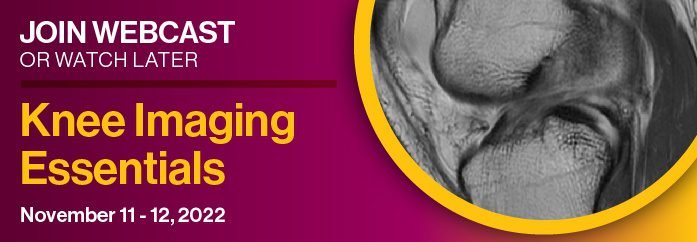
Knee Imaging Essentials 2023 WEBCAST SERIES
$60.00
VIDEO COURSE · Radiology · 2023
Nineteen lectures deliver knee imaging essentials webcast content across two days — covering intricate knee anatomy, imaging protocol optimization, meniscal and cartilage pathology, post-operative evaluation, and pearls and pitfalls from faculty including Brady Huang, Donald Resnick, and William Morrison.
-
19lectures
-
3PDFs
-
2023year
- Lateral and medial supporting structures of the knee
- Menisci and cartilage imaging with tricks and tips
- Post-operative ligament and meniscus evaluation
- Case-based review and hot topics in knee MRI
Knee imaging essentials — 19 lectures on anatomy, protocols, and pathology
The knee imaging essentials webcast provides an update in knee imaging strategies through 19 lectures spread across two days. Faculty share personal tricks and tips for knee image acquisition and interpretation, covering the intricate anatomical structures of the knee, optimized imaging protocols, MRI for sports-related injuries, and pre- and post-operative image characteristics. Brady Huang, Donald Resnick, William Morrison, and other leading faculty deliver content designed for practicing radiologists who supervise and interpret musculoskeletal MRI.
-
19lectures
-
3PDFs
-
2023year
Inside this resource
Anatomy and ligament imaging
Friday’s sessions focus on the anatomic structures that form the foundation of knee MRI interpretation. Brady Huang covers the lateral supporting structures, postoperative ligaments with emphasis on the ACL, and the posterior cruciate ligament. Donald Resnick addresses the medial supporting structures, and a dedicated ACL session rounds out the anatomy-focused day. Understanding these structures at a granular level is what separates a confident knee MRI interpretation from one hedged with unnecessary uncertainty.
Menisci, cartilage, and protocol optimization
Saturday morning tackles the imaging of menisci and cartilage — the structures that generate the most clinical questions. William Morrison leads with tricks and tips for meniscal imaging, John Crues provides a knee MRI update, and Garry Gold addresses menisci and cartilage together. Morrison also covers cartilage and marrow, while John Reeder addresses the pitfalls that can trap even experienced readers.
Case reviews and post-operative evaluation
The program closes with Adam Zoga on knee nuances and nuisances, Lawrence White on the post-operative meniscus, Robert Boutin on case-based review, and Morrison on hot topics in knee MRI. These sessions bridge the gap between pattern recognition and clinical decision-making — teaching you how to report findings in a way that directly influences surgical planning and patient management.
Who this serves
- MSK radiologists — focused knee MRI updates from leading faculty in an efficient two-day format
- Radiology residents — structured learning path from anatomy through pathology and post-operative evaluation
- Referring physicians — understanding what radiologists see on knee MRI to improve clinical correlation
The chart
| Format | 19 video lectures + 3 PDFs |
|---|---|
| Duration | 2 days (November 11–12, 2022) |
| Publisher | Educational Symposia |
| Department | Orthopedics |
| Delivery | Instant download after checkout · lifetime re-access |
Verified original fileInstant downloadEncrypted checkout
5 reviews for Knee Imaging Essentials 2023 WEBCAST SERIES
Add a review
Related products


Orthopedics

Kathleen W. –
Well-organized two-day course covering the full spectrum of knee imaging. The menisci and cartilage sessions are particularly practical. I would have liked more case material but the lecture quality is excellent.
David K. –
Strong faculty delivering focused content on knee imaging. The post-operative evaluation sessions are particularly relevant for my practice. The two-day format is efficient and well-paced.
Robert M. –
Brady Huang and Donald Resnick on knee anatomy in a single course — doesn’t get better than this. The Friday sessions on supporting structures changed how I approach knee MRI. The pitfalls session by Reeder is essential viewing.
Jennifer H. –
Outstanding webcast from Educational Symposia. The case-based review and hot topics sessions bring together the anatomy and pathology covered earlier, creating a complete learning arc. Highly recommended for anyone reading knee MRI.
Steven L. –
The knee imaging essentials live up to their name — these are truly the foundational skills every radiologist interpreting knee MRI needs. Morrison’s meniscal tricks and tips session alone is worth the investment.