
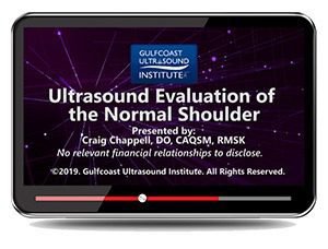
Gulfcoast: Ultrasound Evaluation of the Normal Shoulder

$15.00
VIDEO COURSE · Gulfcoast Ultrasound Institute
This normal shoulder ultrasound evaluation training provides an introduction to shoulder anatomy, probe control, and the sonographic characteristics of normal structures — covering scanning protocol, points of contact, and minor pathology recognition with practical demonstration.
-
7topic areas
- Shoulder anatomy with normal sonographic characteristics
- Probe control and points of contact for systematic scanning
- Scanning protocol for comprehensive shoulder evaluation
- Minor pathology recognition and practical scan demonstration
Normal shoulder ultrasound evaluation for anatomy and scanning technique
This normal shoulder ultrasound evaluation training from Gulfcoast introduces shoulder anatomy, scanning technique, and the sonographic characteristics of normal structures. Seven topic areas establish the baseline knowledge for shoulder assessment — probe control, points of contact, systematic protocol, and normal sonographic appearance — with the minor pathology recognition that helps clinicians distinguish normal variation from early disease.
Inside this resource
Shoulder anatomy and normal sonographic characteristics
The course opens with shoulder anatomy as seen on ultrasound — the rotator cuff tendons, biceps tendon, subacromial bursa, and glenohumeral joint. Normal sonographic characteristics are presented for each structure, establishing the appearance that serves as the reference standard for recognising pathological findings in clinical practice.
Probe control and scanning technique
Probe control and points of contact are demonstrated for each shoulder structure — how transducer pressure, angle, and position affect the image. The scanning protocol organises these techniques into a systematic approach that ensures complete shoulder evaluation, covering the structures from the biceps tendon through the posterior rotator cuff.
Minor pathology and practical scan demonstration
A minor pathology module addresses the early or subtle findings that may appear during a normal shoulder evaluation — small effusions, mild tendinopathy, and the variants that require recognition to avoid misinterpretation. The practical scan demonstration integrates all elements into a reproducible shoulder ultrasound protocol.
Who this serves
- Sonographers new to shoulder MSK scanning — build foundational anatomy knowledge with systematic protocol training
- Radiology and sports medicine trainees — develop shoulder ultrasound technique with structured probe control and protocol demonstration
Physicians learning shoulder ultrasound — establish normal scanning competency before advancing to pathology evaluation
Before you buy
Does this cover shoulder pathology evaluation?
Focused on normal anatomy, scanning technique, and minor pathology recognition — the foundational knowledge before advancing to dedicated shoulder pathology courses.
Is there a companion pathology course?
Yes — the Gulfcoast Shoulder Pathology course covers rotator cuff tears, tendinosis, and diagnostic criteria for commonly seen shoulder conditions.
The chart
| Format | Video lecture (MP4) |
|---|---|
| Publisher | Gulfcoast Ultrasound Institute |
| Department | Gulfcoast Ultrasound Institute |
| Delivery | Instant download after checkout · lifetime re-access |
Verified original fileInstant downloadEncrypted checkout
6 reviews for Gulfcoast: Ultrasound Evaluation of the Normal Shoulder
Add a review
Related products
Gulfcoast Ultrasound Institute
GCUS Introduction to Transcranial Doppler Ultrasound 2019 (VIDEOS)
Gulfcoast Ultrasound Institute
Advanced Musculoskeletal (MSK) Ultrasound Applications 2020 (VIDEOS)
Gulfcoast Ultrasound Institute
Gulfcoast Ultrasound Institute
Gulfcoast Advanced MSK Ultrasound-Guided Procedures for Rehabilitation Medicine
Gulfcoast Ultrasound Institute
AIUM VIDEO HD Musculoskeletal Ultrasound of Sports Injuries and Hernias
Gulfcoast Ultrasound Institute
AIUM Prenatal Ultrasound Abnormalities of the Chest, Abdomen, and Pelvis With Postnatal Correlation

Gulfcoast Ultrasound Institute

Gulfcoast Ultrasound Institute
Lynn C. –
Downloaded alongside the pathology course. Starting with this normal baseline made the shoulder pathology material significantly easier to understand.
James R. –
Excellent foundation for shoulder ultrasound. The probe control and points of contact demonstrations are immediately applicable — helps build scanning confidence early.
drtomh –
Practical for learning shoulder scanning technique. The minor pathology recognition section adds value by showing what normal variation looks like.
Hannah M. –
Good introduction to normal shoulder anatomy. The systematic scanning protocol approach ensures you do not miss structures during evaluation.
Aisha B. –
Useful as a normal anatomy baseline. Would have appreciated more scan demonstration time — the seven topics cover a lot of ground quickly.
Carlos M. –
Well-structured shoulder scanning introduction. The probe control module is particularly well taught — transducer technique makes a real difference in image quality.