

DentistryOnline courseGnathology
Diagnosing the TMJ on CBCT and MRI — Lukas Lassmann links vertical dimension, joint disorders and centric relation
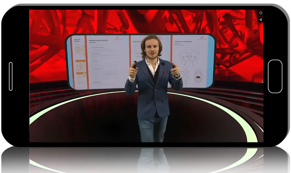
Sound occlusal decisions rest on reading the joint correctly. TMJ radiology and occlusion: vertical dimension, intra-articular disorders, centric relation is Lukas Lassmann’s detailed method for doing that, over six hours and four lessons: how vertical dimension drives TMJ pathology, basic and advanced imaging on pantomograph, CBCT and MRI, and the clinical role of centric relation.
- 4lessons
- 6 h 15 minof video
- 3modalities read
What you’ll be able to do
- Decide when and how much to raise vertical dimension
- Assess condyle position on pantomograph and CBCT
- Distinguish arthritis, arthrosis and resorption radiographically
- Read disc displacement and effusion on MRI
- Establish and question centric relation in real cases
Inside the course
Vertical dimension in depth
The opening lesson connects VDO to joint health, worn dentitions and the cervical spine, and shows how to set a deprogrammer platform height using a modified digital smile design protocol.
From basic to advanced imaging
Two lessons build joint radiology from pantomograph and condyle assessment through cortical erosion, degeneration, resorption and the full MRI picture of disc position and effusion.
Centric relation in practice
The final lesson debates centric slide, long centric, adapted centric posture and the manoeuvres to establish position, ending with when an intervention is truly needed.
About Lukas Lassmann
He lectures internationally, belongs to the Polish Academy of Aesthetic Dentistry and founded a series of advanced clinical courses.
Built for
- Restorative dentistsConfidence to raise occlusion on solid joint evidence.
- OrthodontistsRadiographic reading of joint form and position.
- TMD cliniciansA structured CBCT and MRI interpretation method.
Curriculum
Lesson 1: Vertical dimension of occlusion (1 h 14 min) 13 topics
- When and how much to increase VDO
- Vertical dimension of occlusion explained
- Increasing VDO in excessive tooth wear
- Full mouth reconstruction and TMD
- The 1-1,3-2-3 relation when increasing VDO
- What dentoalveolar compensation is
- Effect of raising VDO on cervical spine and trigeminal pain
- Cephalometric analysis of cervical spine and hyoid position
- Muscle balance as the key to raising occlusion
- Basic photo protocol for aesthetic planning
- Setting deprogrammer platform height with a modified DSD protocol
- DSD in patients with significant tooth abrasion
- The VDO checklist
Lesson 2: TMJ radiology, basic (54 min) 7 topics
- Anamnesis vitae and morbi
- Two steps before ordering radiographs
- Analysis of pantomographic images
- Are 2D images suitable for the TMJ?
- Condyle hypermobility on radiographs
- Structure of the TMJ in radiology
- Condyle position assessed on CBCT
Lesson 3: TMJ radiology, advanced (1 h 43 min) 13 topics
- Why the 4/7 position did not fulfil its role
- Cortical bone erosion on CBCT and how to assess it
- Joint degeneration on CBCT: arthritis versus arthrosis
- Idiopathic condyle resorption and sudden open bite
- Subcortical cysts, ankylosis and developmental defects
- Cephalometry for respiratory disorders
- The hyoid triangle and cervical vertebrae on cephalometry
- MRI normal anatomy relevant to TMD
- Disc displacement with and without reduction
- Analysing joint mobility on MRI
- Joint effusion and the double-disc image
- Why MRI outperforms ultrasound
- Condyle stabilisation seen on MRI
Lesson 4: Clinical application of centric relation (1 h 47 min) 14 topics
- When condyle positioning on MRI and CBCT makes sense
- 4/7 and rearmost positions: why used and abandoned
- Centric slide: physiology or mistake?
- Constricted chewing pattern and long centric
- Rotation versus translation
- Defining the centric relation
- Adapted centric posture and when to intervene
- Muscle vectors of the stomatognathic system
- Establishing centric relation with deprogrammers and manoeuvres
- Triple centric relation and articular fossa position
- Forward position: splint, functional orthodontics, apnea
- Neuromuscular position: principles and doubts
- Which position to choose and when
- Does an incorrect joint position guarantee TMD?
Before you buy
Do I need imaging experience?
It builds from basic pantomograph reading to advanced CBCT and MRI, so prior radiology is helpful but not assumed.
Is it clinical or purely diagnostic?
Both. Imaging interpretation is tied throughout to vertical dimension decisions and centric relation in restorative and orthodontic care.
Do I get CME credit or a certificate?
No. Clinicapedia provides the educational content only — no CME or CE credits, MOC points or certificates of completion are included with this product.
Specifications
| Format | Online video course |
|---|---|
| Instructor | Lukas Lassmann |
| Contents | 4 lessons |
| Duration | 6 h 15 min |
| Access | Lifetime, self-paced |
| Department | Dentistry |
| Delivery | Instant download · lifetime re-download |
Related products

Dentistry



Anesthesiology