
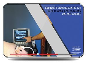
Advanced Musculoskeletal (MSK) Ultrasound Applications 2020 (VIDEOS)
$45.00
VIDEO COURSE · Gulfcoast Ultrasound Institute · 2020
The advanced MSK ultrasound course from Gulfcoast delivers eight recorded lectures on dynamic joint imaging, peripheral nerve assessment, and post-operative evaluation — built for sonographers and physicians expanding their musculoskeletal diagnostic range.
-
8video lectures
-
3.32 GBlibrary size
-
8topics covered
-
2020edition
- Dynamic maneuvers for hip, ankle, and post-operative joint pathology
- Peripheral nerve ultrasound and guided injection protocols
- Spine imaging with lumbar and cervical injection techniques
- MSK tumor and mass characterization with case examples
Advanced MSK ultrasound applications for dynamic joint and nerve imaging
This advanced MSK ultrasound course equips clinicians with scanning protocols and diagnostic criteria for musculoskeletal pathologies that demand dynamic maneuvers and specialized technique. Eight lectures cover hip evaluation, foot and ankle ligament integrity, post-operative joint assessment, peripheral nerve imaging, spine ultrasound, and tumor characterization — all taught through real case examples and step-by-step demonstrations.
-
8video lectures
-
3.32 GBlibrary size
-
8topic modules
-
2020release year
Inside this resource
Dynamic joint assessment and post-operative evaluation
The course opens with dynamic MSK imaging — joint movement, patient positioning, and muscle contraction maneuvers that reveal pathology static scans miss. Hip anatomy and pathology receive dedicated coverage, along with post-operative joint evaluation using diagnostic criteria specific to surgical implants and repair sites.
Foot, ankle, and peripheral nerve applications
Lectures walk through the calcaneofibular, anterior inferior tibiofibular, and anterior talofibular ligaments with dynamic ultrasound examination. Medial ankle tendons, ligaments, and neurovascular structures are covered alongside scanning protocols for peripheral nerve ultrasound and indications for guided nerve injections in MSK practice.
Spine imaging and tumor characterization
The spine module addresses lumbar and cervical anatomy best evaluated with ultrasound, paired with injection technique demonstrations for spine interventions. A final lecture tackles MSK tumors and masses — their ultrasound characteristics, pseudotumor differentiation, and the patterns that guide clinical decision-making.
Who this serves
- Sonographers — expand into advanced MSK scanning with protocols for dynamic maneuvers and nerve imaging
- Physicians — build interpretation skills for complex joint, post-operative, and spine applications
- Mid-level providers — strengthen diagnostic accuracy for musculoskeletal ultrasound examinations
Faculty & syllabus
Topics covered 8 sessions
- Dynamic Musculoskeletal Ultrasound Imaging
- Advanced Ultrasound Techniques for the Foot and Ankle
- Ultrasound Evaluation of the Hip
- Musculoskeletal Ultrasound for Post-Operative Applications
- Ultrasound Evaluation of Peripheral Nerves and Focal Neuropathies
- Ultrasound-Guided Peripheral Nerve Injections in an MSK Practice
- Ultrasound Evaluation of the Spine and Injection Techniques
- MSK Ultrasound of Tumors and Masses
Before you buy
What is the file format?
Eight MP4 video lectures totaling 3.32 GB, downloadable after purchase with lifetime re-access.
Does this cover interventional techniques?
Yes — the course addresses both diagnostic evaluation of dynamic pathology and ultrasound-guided injection techniques for peripheral nerves and the spine.
The chart
| Format | 8 video lectures (MP4) |
|---|---|
| File Size | 3.32 GB |
| Year | 2020 |
| Publisher | Gulfcoast Ultrasound Institute |
| Department | Gulfcoast Ultrasound Institute |
| Delivery | Instant download after checkout · lifetime re-access |
Verified original fileInstant downloadEncrypted checkout
6 reviews for Advanced Musculoskeletal (MSK) Ultrasound Applications 2020 (VIDEOS)
Add a review
Related products

Gulfcoast Ultrasound Institute
Gulfcoast Ultrasound Institute
AIUM VIDEO HD Musculoskeletal Ultrasound of Sports Injuries and Hernias

Gulfcoast Ultrasound Institute
GCUS Introduction to Carotid Duplex/Color Flow Ultrasound 2022 (VIDEOS)

Gulfcoast Ultrasound Institute
Gulfcoast Ultrasound Institute
Gulfcoast Ultrasound Institute
Gulfcoast Use of Ultrasound in Rheumatology Applications (Videos+PDFs)
Gulfcoast Ultrasound Institute
GCUS Abdominal and Primary Care Ultrasound 2022 (Gulfcoast Ultrasound Institute) (Videos) 2022
Gulfcoast Ultrasound Institute
Gulfcoast Upper Extremity Musculoskeletal Ultrasound Protocols (Videos)
James R. –
The tumor and mass characterization lectures gave me patterns I now reference regularly. Well-organized and worth the price for the spine module alone.
M. Alvarez –
Solid coverage of foot and ankle ligaments with dynamic assessment. The peripheral nerve section is well done. Would have liked a few more injection case examples.
Priya –
Good overview of advanced MSK topics. The dynamic imaging section stands out from introductory courses. File size is reasonable at 3.32 GB.
drtomh –
Downloaded for the spine and tumor lectures. Both exceeded my expectations — the lumbar injection technique demos are especially practical for daily use.
Lynn C. –
Content quality is solid but I wanted more case studies showing pathology variation across different joints. Eight lectures is a tight scope for the price.
Sarah K. –
The hip evaluation module was exactly what I needed. Dynamic maneuvers are demonstrated clearly and the post-operative cases tie everything together nicely.