
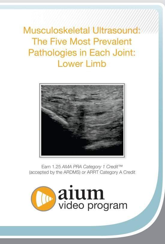
AIUM MSK Ultrasound: The Five Most Prevalent Pathologies in Each Joint: Lower Limb


$15.00
VIDEO COURSE · AIUM · MSK Lower Limb
This lower limb MSK pathologies course covers the five most prevalent ultrasound findings in the knee, hip, and ankle — including the sonographic appearance of commonly encountered pathology and the role of ultrasound-guided procedures in treatment of these conditions.
-
3joint regions
- Five most common pathologies in knee, hip, and ankle
- Sonographic appearance and diagnostic criteria
- Ultrasound-guided procedures for treatment
- Hip pain categorisation: anterior, lateral, and posterior
Lower limb MSK ultrasound: five pathologies per joint in knee, hip, and ankle
This lower limb MSK pathologies course from the AIUM systematically covers the five most prevalent ultrasound findings in three lower limb joints — knee, hip, and ankle. Each joint receives dedicated attention with the sonographic appearance, diagnostic criteria, and the role of ultrasound-guided procedures in managing these pathologies. The course addresses hip pain categorisation by location and the ankle pathologies where ultrasound guidance improves procedural outcomes.
Inside this resource
Knee pathology assessment
The knee module covers the five most common pathologies seen with ultrasound — meniscal tears, ligament injuries, bursitis, Baker cysts, and patellar tendon pathology. Each condition is presented with its sonographic appearance, the scanning approach that reveals the findings, and the clinical correlation that connects imaging to physical examination.
Hip pain evaluation by location
Hip pain is categorised by anatomical location — anterior, lateral, and posterior — with the differential diagnosis that guides further evaluation for each region. Normal hip anatomy serves as the baseline, and the five most prevalent pathologies are presented with their sonographic features and clinical significance.
Ankle and foot pathology with procedural guidance
The ankle module addresses the five most prevalent pathologies — including ligament injuries, tendinopathy, and enthesopathy — and the important role of ultrasound-guided procedures in their treatment. The procedural component demonstrates how sonographic visualisation improves injection accuracy in the ankle and foot.
Who this serves
- Sonographers in MSK practice — build systematic knowledge of lower limb joint pathology with ultrasound
- Physicians performing MSK ultrasound — develop diagnostic and procedural competency for knee, hip, and ankle
- Sports medicine clinicians — strengthen lower limb pathology recognition for athletic injury assessment
Before you buy
Does this cover all three joints equally?
Yes — knee, hip, and ankle each receive dedicated coverage with their five most prevalent pathologies, sonographic appearance, and clinical significance.
Is this a lower-limb-only course?
This volume covers the lower limb (knee, hip, ankle). A companion volume addresses the upper limb (shoulder, hand, wrist).
The chart
| Format | Video lectures (MP4) |
|---|---|
| Publisher | AIUM |
| Department | Gulfcoast Ultrasound Institute |
| Delivery | Instant download after checkout · lifetime re-access |
Verified original fileInstant downloadEncrypted checkout
5 reviews for AIUM MSK Ultrasound: The Five Most Prevalent Pathologies in Each Joint: Lower Limb
Add a review
Related products

Gulfcoast Ultrasound Institute

Gulfcoast Ultrasound Institute

Gulfcoast Ultrasound Institute
GCUS Introduction to Transcranial Doppler Ultrasound 2019 (VIDEOS)

Gulfcoast Ultrasound Institute
Gulfcoast Ultrasound Institute
GCUS Introduction to Peripheral Vascular Ultrasound 2022 (VIDEOS)

Gulfcoast Ultrasound Institute
Gulfcoast Ultrasound Institute
Gulfcoast Advanced MSK Ultrasound-Guided Procedures for Rehabilitation Medicine
Gulfcoast Ultrasound Institute
AIUM VIDEO HD Musculoskeletal Ultrasound of Sports Injuries and Hernias
Wei L. –
Downloaded as a reference for lower limb MSK scanning. The systematic joint-by-joint approach makes it easy to review specific regions as needed.
Marcus J. –
Systematic approach to the five most common pathologies in each lower limb joint. The hip pain categorisation by location is particularly useful for clinical assessment.
Sarah K. –
Practical for MSK scanning practice. Each condition is presented with clear sonographic criteria and the clinical correlation that supports pattern recognition.
James R. –
Good coverage of knee, hip, and ankle pathology. The ankle procedural guidance section demonstrates how ultrasound improves injection accuracy.
drtomh –
Content is solid but I wanted more on diagnostic criteria variations across different pathology grades. The five-per-joint structure works well as an organiser.