
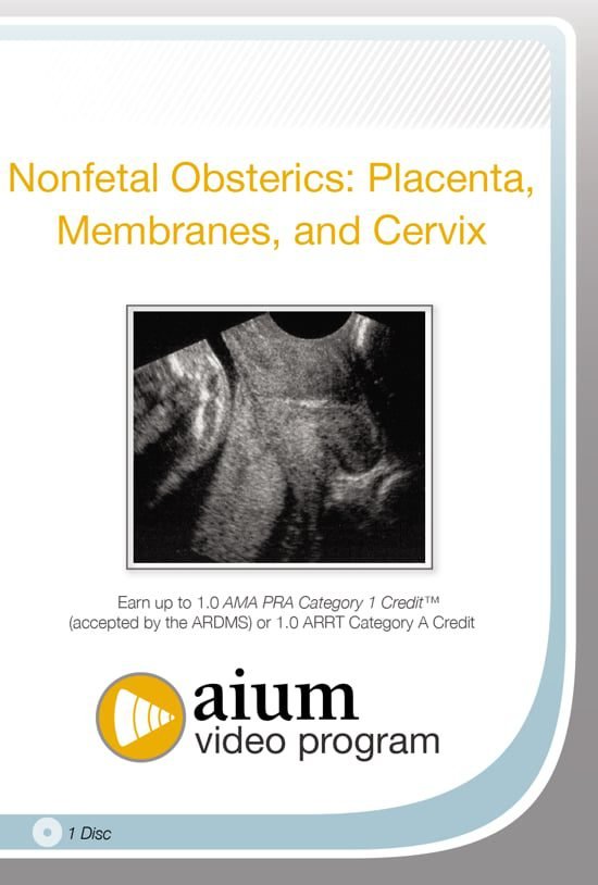
Nonfetal Obstetrics: Placenta, Membranes, and Cervix

$15.00
VIDEO COURSE · AIUM · Nonfetal Obstetrics
This placenta membranes cervix ultrasound course reviews the sonographic evaluation of nonfetal obstetric structures — the placenta, membranes, and cervix — covering the imaging features that inform clinical management during pregnancy without focusing on fetal anatomy itself.
-
1video lecture
- Placental localisation and pathology assessment
- Membrane evaluation for clinical decision-making
- Cervical length and morphology sonography
- Nonfetal obstetric structures in pregnancy management
Placenta, membranes, and cervix ultrasound evaluation in obstetric practice
This placenta membranes cervix ultrasound course from the AIUM reviews the sonographic assessment of the nonfetal structures that influence pregnancy management — the placenta, membranes, and cervix. The training addresses patient care and medical knowledge attributes through a practice-based learning approach, teaching clinicians how to evaluate placental position and pathology, membrane integrity, and cervical morphology.
Inside this resource
Placental evaluation and localisation
The placenta module covers sonographic localisation — determining placental position relative to the internal os, identifying placenta praevia, and assessing for placental abruption and other pathology. The grading system for placental maturity and the Doppler features that assess intervillous blood flow receive attention alongside the grey-scale findings.
Membrane assessment and cervical sonography
Membrane evaluation addresses the sonographic findings relevant to preterm premature rupture of membranes — including the identification of the membranes, assessment of amniotic fluid volume, and the role of ultrasound in confirming membrane status. The cervical module covers length measurement, funneling assessment, and the findings that predict preterm delivery risk.
Clinical integration and management implications
Each section ties the sonographic findings back to clinical management decisions — when cervical cerclage is indicated, how placental position affects delivery planning, and what membrane findings guide admission versus outpatient management. The aim is connecting imaging to the actionable next steps in obstetric care.
Who this serves
- Obstetric sonographers — develop systematic evaluation skills for nonfetal obstetric structures
- Obstetricians — refine ultrasound assessment of placenta, membranes, and cervix for clinical decision-making
- Trainees in maternal-fetal medicine — build foundational knowledge of nonfetal obstetric ultrasound
Before you buy
Does this cover fetal anatomy assessment?
No — this course focuses specifically on the nonfetal structures: placenta, membranes, and cervix. Fetal anatomy evaluation is addressed in other AIUM tutorials.
What clinical scenarios does this address?
Placental localisation and pathology, membrane integrity assessment, and cervical evaluation for preterm delivery risk — all with management implications for obstetric practice.
The chart
| Format | Video lecture (MP4) |
|---|---|
| Publisher | AIUM |
| Department | Gulfcoast Ultrasound Institute |
| Delivery | Instant download after checkout · lifetime re-access |
Verified original fileInstant downloadEncrypted checkout
5 reviews for Nonfetal Obstetrics: Placenta, Membranes, and Cervix
Add a review
Related products
Gulfcoast Ultrasound Institute
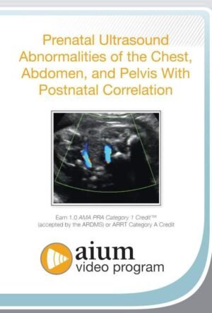
AIUM Prenatal Ultrasound Abnormalities of the Chest, Abdomen, and Pelvis With Postnatal Correlation

Gulfcoast Ultrasound Institute
GCUS Introduction to Carotid Duplex/Color Flow Ultrasound 2022 (VIDEOS)
Gulfcoast Ultrasound Institute
Gulfcoast Ultrasound Institute
Gulfcoast Ultrasound Institute
Gulfcoast Upper Extremity Musculoskeletal Ultrasound Protocols (Videos)

Gulfcoast Ultrasound Institute

Gulfcoast Ultrasound Institute
Gulfcoast Ultrasound Institute
M. Alvarez –
Downloaded as a refresher before an MFM rotation. The systematic approach to nonfetal obstetric evaluation is exactly what I needed for clinical practice.
Hannah M. –
Finally a focused resource on placenta and cervix assessment. The cervical length measurement technique and funneling identification are clearly demonstrated.
Wei L. –
Useful for understanding the management implications of ultrasound findings. The clinical integration approach connects imaging to decision-making effectively.
Natalie F. –
Good overview of nonfetal obstetric structures. The placental localisation module with praevia identification is clinically relevant and well-presented.
Carlos M. –
Solid content but brief. The membrane assessment section felt shorter than the placenta module — would have appreciated more on preterm rupture evaluation.