
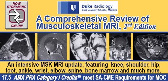
Duke Radiology: A Comprehensive Review of Musculoskeletal MRI, 2nd Edition Agenda

$10.00
VIDEO COURSE · Orthopedics · 2018
Duke Radiology’s musculoskeletal MRI review course delivers an 11-session comprehensive curriculum covering knee anatomy and protocols, ligaments and menisci, hip and pelvis, shoulder, wrist and hand, foot and ankle, elbow, lumbar spine, and bone marrow tumors — taught by Clyde A. Helms, MD and James B. Vogler III, MD.
-
11sessions
-
2faculty
-
2018year
- Systematic joint-by-joint approach to MSK MRI interpretation
- Knee protocols, meniscal tears, cruciate and collateral ligament evaluation
- Hip and pelvis osseous abnormalities, labral tears, FAI, and arthroplasty MRI
- Lumbar spine disc disease, stenosis, spondylolysis, and bone marrow pathology
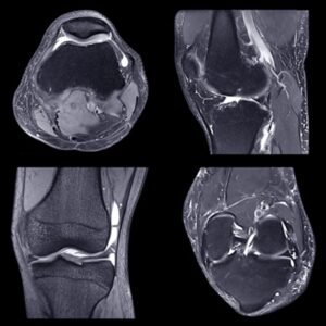
Duke Radiology comprehensive musculoskeletal MRI — 11 sessions from knee to spine
The musculoskeletal MRI review from Duke Radiology, now in its second edition, provides a structured, joint-by-joint approach to MSK MRI interpretation across 11 sessions. Clyde A. Helms, MD and James B. Vogler III, MD — both experienced musculoskeletal imagers — guide you through normal anatomy, protocols, pathological findings, and diagnostic pitfalls for every major joint. The curriculum is designed for radiologists, orthopaedic surgeons, and imaging fellows who need a systematic framework for reading MSK MRI studies.
-
11sessions
-
2faculty
-
2018year
Inside this resource
Knee MRI — anatomy, menisci, and ligaments
The course opens with two dedicated knee sessions. Helms covers anatomy and protocols before moving through meniscal tears, discoid menisci, cysts, cruciate and collateral ligaments, patella, bursae, and cartilage. The systematic approach builds from normal imaging characteristics to the specific tear patterns and ligamentous injuries that dominate daily MSK practice.
Hip, pelvis, shoulder, and wrist
Vogler addresses hip and pelvis osseous abnormalities, tendon pathology, labral tears, FAI, and MRI of hip arthroplasties. Helms returns for two shoulder sessions covering anatomy, rotator cuff tears, labral pathology, glenohumeral instability, and normal variants. The wrist and hand session with Vogler examines technique, osseous abnormalities, ligaments, TFCC, tendons, nerves, and masses — a joint frequently underrepresented in MSK curricula.
Foot and ankle, elbow, spine, and tumors
Three foot and ankle sessions with Helms cover tendons in three parts, ligaments, tumors, bony abnormalities, and osteochondral injuries. Vogler addresses elbow ligament injuries and muscle-tendon abnormalities. The lumbar spine session covers disc disease, mimics, stenosis, spondylolysis, and spondylolisthesis. The final session tackles primary bone and soft tissue tumors, hemorrhage, intra-articular masses, and bone marrow evaluation — closing with a Q&A that ties the curriculum together.
Who this serves
- Radiologists and imaging fellows — systematic MSK MRI review for clinical practice or CAQ preparation
- Orthopaedic surgeons — deeper understanding of MRI interpretation for surgical planning
- Emergency medicine and sports medicine clinicians — improved recognition of meniscal, ligamentous, and tendon pathology on MSK studies
Faculty & syllabus
Faculty 2 speakers
- Clyde A. Helms, MD — knee, shoulder, foot/ankle, lumbar spine, tumors, bone marrow
- James B. Vogler III, MD — hip/pelvis, wrist/hand, elbow, lumbar spine
Sessions covered 11 sessions
- Knee anatomy, protocols, and menisci
- Knee ligaments, patella, bursae, and cartilage
- Pelvis and hips
- Shoulder I — anatomy, pathology, rotator cuff
- Shoulder II — labrum, instability, variants
- Wrist and hand
- Foot and ankle I — tendons
- Foot and ankle II — ligaments, tumors, injuries
- Elbow — ligament injuries, muscle and tendon
- Lumbar spine — disc disease, stenosis, spondylolysis
- Tumors and bone marrow
Before you buy
Is this the same as the Duke Radiology Annual Overview?
No — this is a dedicated 2nd-edition musculoskeletal MRI course focused exclusively on MSK imaging, not a general radiology overview.
Which anatomical regions does this review address?
Knee, hip and pelvis, shoulder, wrist and hand, foot and ankle, elbow, and lumbar spine — plus a dedicated tumors and bone marrow session.
The chart
| Format | Video lectures |
|---|---|
| Publisher | Duke Radiology |
| Department | Orthopedics, Radiology |
| Delivery | Instant download after checkout · lifetime re-access |
Verified original fileInstant downloadEncrypted checkout
5 reviews for Duke Radiology: A Comprehensive Review of Musculoskeletal MRI, 2nd Edition Agenda
Add a review
Related products


Orthopedics



Omar F. –
Used this for MSK MRI CAQ prep and it covered nearly every joint in a logical sequence. The hip/pelvis session with Dr. Vogler on FAI and arthroplasty MRI was particularly relevant to my practice.
Richard P. –
Dr. Helms has a gift for making MSK MRI interpretation feel systematic. The knee and shoulder sessions are the best I have taken — each finding is tied to a clear diagnostic framework.
Lisa D. –
Good comprehensive review with a focused MSK scope. The lumbar spine and tumors sessions rounded out the curriculum nicely. Two experienced faculty teaching the whole course gives it consistency.
Nina K. –
Strong joint-by-joint review. The wrist and hand session filled a gap I did not realize I had. The foot and ankle tendon lectures are thorough. Would have loved more case examples but the anatomy coverage is solid.
Peter W. –
The content is solid but the production quality is a bit dated. The anatomy and pathology teaching by both faculty is excellent — the knee menisci and shoulder rotator cuff sessions are worth revisiting repeatedly.