
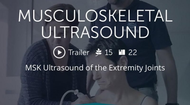
123sonography Musculoskeletal Ultrasound BachelorClass

$40.00
VIDEO COURSE · Orthopedics · 2022
123sonography musculoskeletal ultrasound basics course delivers 174 short videos and micro-learning modules across 22 chapters — covering anatomical tissue recognition, scanning techniques, protocol positions, and pathology checklists for the shoulder, elbow, wrist, hip, knee, and ankle.
-
174videos
-
18.3 GBsize
-
22chapters
-
2022year
- Micro-learning format: short videos for focused, digestible study sessions
- Joint-by-joint anatomy, protocol positions, and pathology for six major joints
- Qualitative scanning techniques and anatomical tissue recognition
- Pathology checklist for systematic MSK ultrasound assessment
123sonography musculoskeletal ultrasound BachelorClass — 174 short videos for daily practice
The 123sonography musculoskeletal ultrasound basics course BachelorClass is an exclusive teaching program built around 174 short videos and micro-learning moments. Designed to teach the fundamentals of MSK ultrasound from the ground up, the curriculum covers anatomical tissue recognition, qualitative scanning techniques, and joint-by-joint anatomy, protocol positions, and pathology for six major joints — shoulder, elbow, wrist, hip, knee, and ankle. The short-form video format lets you study specific topics in focused sessions rather than sitting through long lectures.
-
174short videos
-
18.3 GBsize
-
22chapters
-
2022year
Inside this resource
Foundations — tissue recognition and scanning technique
The opening chapters establish the basics: understanding anatomical tissues on ultrasound — how tendons, ligaments, muscles, cartilage, bone, and fluid appear sonographically. Qualitative scanning covers probe selection, gain optimization, and the technique principles that underpin every joint examination. This foundation ensures that the joint-specific chapters that follow build on a consistent skill set.
Joint-by-joint anatomy and protocol positions
Each of the six major joints — shoulder, elbow, wrist, hip, knee, and ankle — gets three dedicated chapters: anatomy, protocol positions, and pathology. The anatomy chapters teach normal sonographic appearance. Protocol positions cover the standardized scanning sequences for each joint — which views to obtain, in what order, and what structures to identify at each station. This protocol-driven approach mirrors how MSK ultrasound departments organize their examinations.
Pathology checklist and clinical application
The final chapters provide a pathology checklist — a systematic framework for identifying abnormal findings during MSK ultrasound examinations. This checklist approach ensures you do not miss findings when scanning under time pressure. Combined with the anatomy and protocol chapters, the curriculum gives you both the knowledge base and the practical workflow to perform competent MSK ultrasound examinations in clinical practice.
Who this serves
- Ultrasound beginners — structured entry point into MSK sonography with short, digestible video modules
- Clinicians adding MSK ultrasound — protocol-driven training for primary care, sports medicine, and rheumatology
- Sonographers cross-training into MSK — systematic anatomy and pathology review for new MSK departments
Before you buy
How long are the individual videos?
The program uses short-form micro-learning videos — each focused on a specific topic, technique, or structure, designed for brief study sessions.
Which body regions are covered in the curriculum?
Six major joints: shoulder, elbow, wrist, hip, knee, and ankle — each with anatomy, protocol positions, and pathology chapters.
The chart
| Format | 174 short videos (.mp4) |
|---|---|
| Publisher | 123sonography |
| Department | Orthopedics, Radiology |
| Delivery | Instant download after checkout · lifetime re-access |
Verified original fileInstant downloadEncrypted checkout
5 reviews for 123sonography Musculoskeletal Ultrasound BachelorClass
Add a review
Related products




Maria L. –
The micro-learning format is perfect for busy clinicians. I watch a few videos between patients and the joint-by-joint protocol approach builds systematically. The pathology checklists are a great reference.
Robert F. –
Good foundational course with a practical protocol-driven approach. The ankle and wrist modules are thorough and often overlooked in MSK ultrasound programs. Works well for self-paced learning.
James C. –
Solid introduction to MSK ultrasound. The tissue recognition chapters laid a good foundation before moving to joint-specific protocols. At 174 videos and 18.3 GB, it is a large collection but well organized.
Laura M. –
The basics are well covered but more advanced users may find this too introductory. For beginners, the short video format and systematic approach make it a solid entry point into MSK ultrasound.
Sarah K. –
Exactly what I needed as a complete beginner in MSK sonography. The protocol positions for each joint give me a reproducible scanning sequence. The shoulder and knee pathology chapters are particularly well done.