
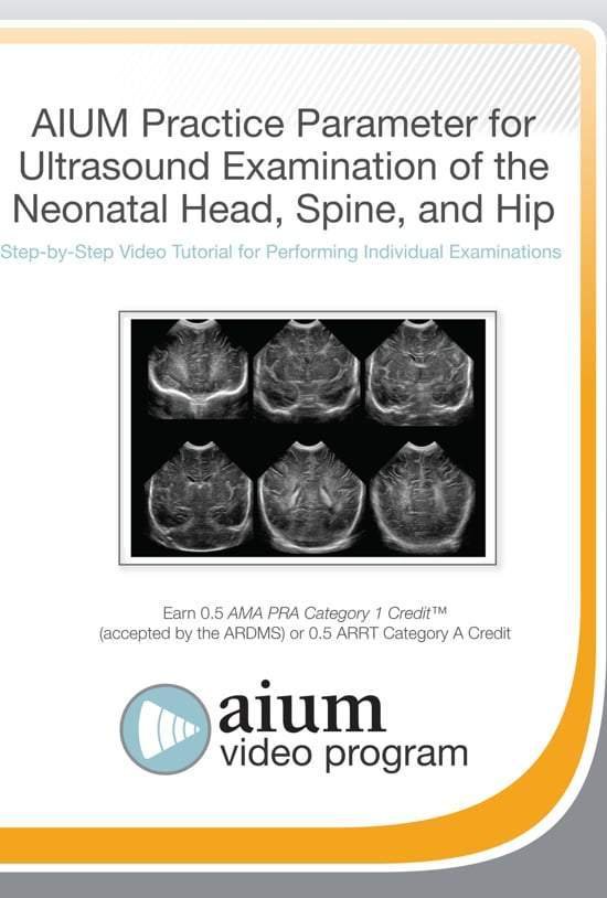
AIUM Practice Parameteres for Neonatal Head, Spine, and Hip
$15.00
VIDEO COURSE · AIUM · Neonatal Imaging
This neonatal head spine hip ultrasound tutorial demonstrates the techniques for performing neonatal ultrasound examinations of the brain, spine, and hip — covering the normal sonographic appearance across three practice parameters for neurosonography, spinal imaging, and developmental dysplasia of the hip.
-
3anatomical regions
- Normal sonographic appearance of the neonatal brain
- Neonatal and infant spine ultrasound technique
- Developmental dysplasia of the hip assessment
- Three AIUM practice parameters in a single tutorial
Neonatal head, spine, and hip ultrasound practice parameter tutorial
This neonatal head spine hip ultrasound tutorial from the AIUM demonstrates the scanning techniques for three critical neonatal applications — neurosonography of the brain, spinal ultrasound in neonates and infants, and assessment for developmental dysplasia of the hip. Each section follows the corresponding AIUM practice parameter, teaching the normal sonographic appearance that serves as the baseline for identifying pathology.
Inside this resource
Neonatal brain neurosonography
The brain section covers the normal sonographic anatomy of the neonatal cranial structures — ventricles, parenchyma, germinal matrix, and cerebellum. The tutorial demonstrates anterior fontanelle and mastoid fontanelle scanning windows, with the standardised planes recommended by the AIUM practice parameter for neurosonography in neonates and infants.
Neonatal and infant spine evaluation
The spine module addresses the sonographic appearance of the normal neonatal and infant spine — vertebral bodies, spinal canal, conus medullaris, and the posterior elements. Scanning technique through the lumbar and sacral windows is demonstrated, following the AIUM practice parameter for spinal ultrasound examination.
Developmental dysplasia of the hip assessment
The hip section teaches the ultrasound evaluation for developmental dysplasia of the hip — acetabular coverage, femoral head position, and the Graf classification approach. The AIUM practice parameter for detection and assessment of hip dysplasia guides the scanning technique and measurement methodology.
Who this serves
- Neonatal sonographers — align technique with AIUM practice parameters for brain, spine, and hip imaging
- Paediatric radiologists — review normal neonatal sonographic anatomy across three regions
- Neonatologists and paediatricians — understand the ultrasound findings used in routine neonatal assessment
Before you buy
Does this cover pathology or normal anatomy?
The tutorial focuses on normal sonographic appearance — the baseline knowledge needed before identifying pathological findings in neonatal brain, spine, and hip imaging.
Which practice parameters does this follow?
Three AIUM practice parameters: neurosonography in neonates, neonatal spine ultrasound, and detection of developmental dysplasia of the hip.
The chart
| Format | Video tutorial (MP4) |
|---|---|
| Publisher | AIUM |
| Department | Gulfcoast Ultrasound Institute |
| Delivery | Instant download after checkout · lifetime re-access |
Verified original fileInstant downloadEncrypted checkout
5 reviews for AIUM Practice Parameteres for Neonatal Head, Spine, and Hip
Add a review
Related products

Gulfcoast Ultrasound Institute
Gulfcoast Ultrasound Institute
Gulfcoast Advanced Ultrasound Techniques for the Foot and Ankle (Videos+PDFs)
Gulfcoast Ultrasound Institute
GCUS Introduction to Carotid Duplex/Color Flow Ultrasound 2022 (VIDEOS)
Gulfcoast Ultrasound Institute
GCUS Introduction to Peripheral Vascular Ultrasound 2022 (VIDEOS)
Gulfcoast Ultrasound Institute
GCUS Abdominal and Primary Care Ultrasound 2022 (Gulfcoast Ultrasound Institute) (Videos) 2022
Gulfcoast Ultrasound Institute
AIUM VIDEO HD Musculoskeletal Ultrasound of Sports Injuries and Hernias

Gulfcoast Ultrasound Institute

Gulfcoast Ultrasound Institute
Fatima R. –
Exactly what I needed for neonatal scanning competency. The standardised planes for each region are clearly labelled and easy to reproduce.
Sarah K. –
Three AIUM practice parameters in one tutorial — excellent value. The neurosonography section with normal anatomy baseline is well-demonstrated.
Hannah M. –
Downloaded for a neonatal scanning refresher. The normal anatomy emphasis provides a solid foundation before tackling pathological findings.
Natalie F. –
Good overview of neonatal brain, spine, and hip ultrasound. The hip dysplasia assessment with Graf classification is clearly presented.
Raj P. –
Practical content but brief. The spine section felt shorter than the brain and hip modules — would have liked more detail on conus evaluation.