
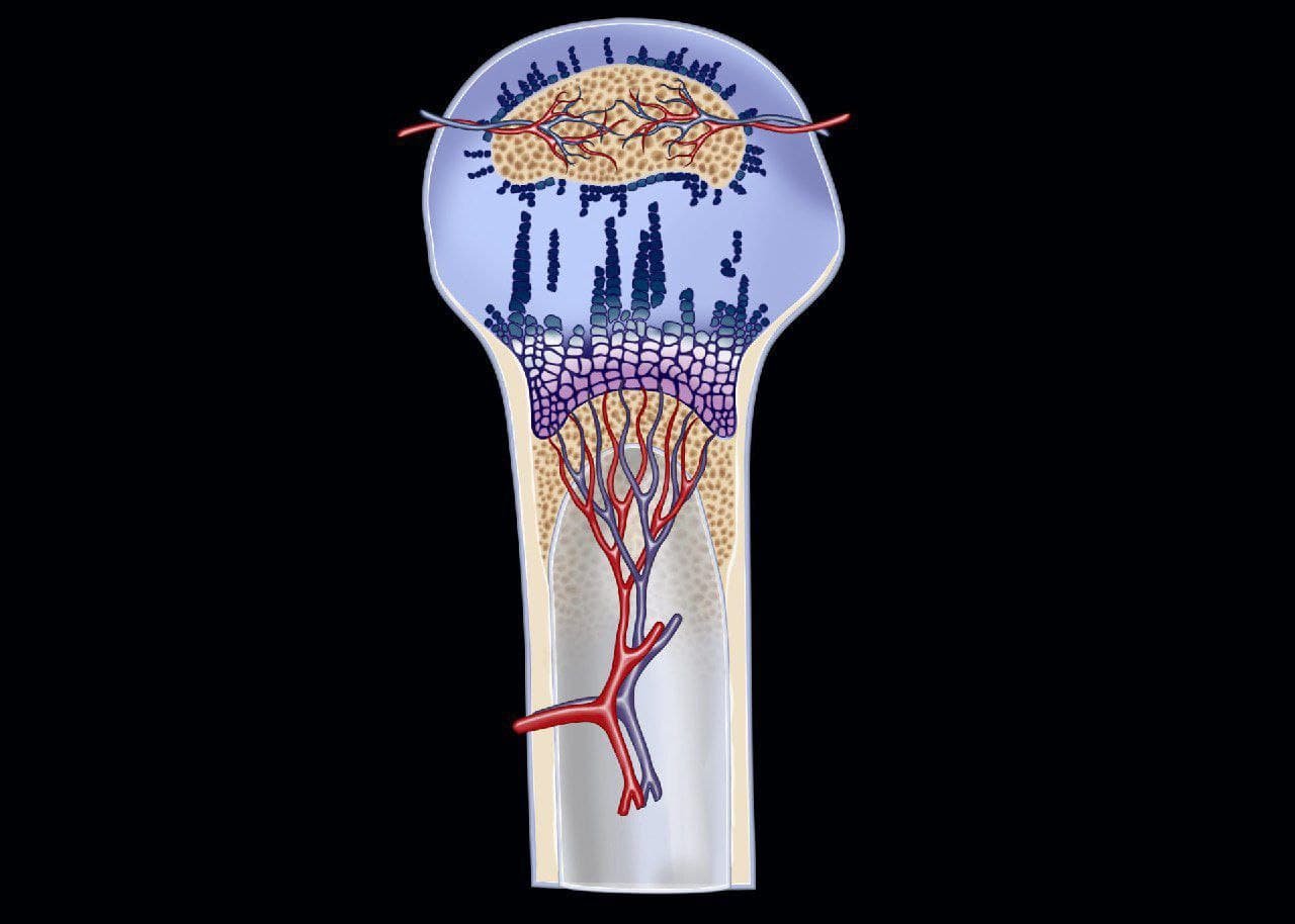
MRIOnline Imaging Mastery Series: Pediatric MSK 2021 (CME VIDEOS)


$25.00
Video Course · Musculoskeletal · 2021
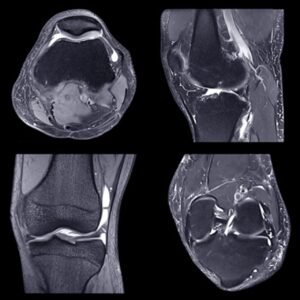
The pediatric MSK MRI mastery series from MRIOnline delivers 71 video lectures by Dr. Thapa on cartilage injury patterns, growth plate pathology, and musculoskeletal abnormalities unique to the pediatric population.
-
71video lectures
-
2021edition
- Covers Salter-Harris fractures, FOPE lesions, and avascular necrosis
- Reviews OCD in the elbow, capitulum, and knee including trochlear variants
- Includes Blount disease, gymnast wrist, and Legg-Calve-Perthes evaluation
- Teaches age-specific cartilage signal patterns and ossification center recognition
71 Lectures on Pediatric Cartilage Injury and Growth Plate Pathology
The pediatric MSK MRI mastery series by MRIOnline features Dr. Thapa presenting 71 lectures on the unique imaging challenges of pediatric musculoskeletal patients. Pediatric patients are not simply small adults — differences range from open growth plates to nuanced ossification center signal intensities, and knowing when a finding is abnormal versus a normal age-variant is central to accurate interpretation.
-
71video lectures
-
2021edition
Inside this resource
Cartilage Anatomy and Age-Specific MRI Appearance
Foundational sessions cover hyaline cartilage anatomy, the physis and calcification centers, epiphyseal cartilage, fibrocartilage, and how cartilage signal changes across different age groups. Lectures on pre-ossification centers and MR appearance of cartilage in different age groups establish the interpretive framework used throughout the pediatric MSK MRI curriculum.
Elbow Pathology and OCD Evaluation
Dedicated sessions address elbow effusion, OCD in the elbow, trochlear OCD on MRI, trochlear OCD on arthrogram, ultrasound-guided arthrogram injection, OCD in the capitulum with loose body, avascular necrosis in the elbow, and the fish tail deformity. Each lecture applies the pediatric MSK MRI framework to a specific elbow diagnosis.
Knee, Hip, and Extremity Diagnoses
The series continues with OCD in the knee and LAME, Legg-Calve-Perthes disease on X-ray, Salter-Harris fractures, FOPE lesions, Blount disease, gymnast wrist, and lymphoma of bone. Each session teaches the learner to formulate definitive diagnoses and limited differentials using appropriate search patterns.
Who it’s for
This pediatric MSK MRI series serves pediatric radiologists, general radiologists who evaluate musculoskeletal cases in children, orthopedic surgeons seeking imaging literacy for growing patients, and radiology residents building foundational competency in pediatric joint and bone imaging.
Specs
| Format | 71 MP4 video lectures |
| Edition | 2021 |
| Delivery | Instant digital download |
| File Size | 3.02 GB |
FAQ
What imaging modalities does the pediatric MSK MRI series cover?
The curriculum focuses primarily on MRI evaluation of pediatric musculoskeletal conditions, with supporting sessions on X-ray findings and ultrasound-guided procedures for elbow arthrogram injection.
Is this suitable for someone new to pediatric MSK MRI?
The series opens with foundational lectures on cartilage anatomy and age-specific MRI appearance before progressing to specific diagnoses, making it accessible for radiologists transitioning into pediatric musculoskeletal imaging.
Does it cover both upper and lower extremity pathology?
Yes. Lectures address elbow conditions including OCD variants and avascular necrosis, knee diagnoses such as LAME and Salter-Harris fractures, hip pathology like Legg-Calve-Perthes disease, and wrist findings including gymnast wrist.
5 reviews for MRIOnline Imaging Mastery Series: Pediatric MSK 2021 (CME VIDEOS)
Add a review
Related products

Pulmonary /Respiratory
ARRS Lung Cancer Screening A Comprehensive Guide Online Course
Pulmonary /Respiratory

Natalie R. –
Dr. Thapa breaks down cartilage anatomy in a way that finally made FOPE lesions and pre-ossification centers click for me. The 71 lectures progress logically from anatomy through specific diagnoses. An essential reference for anyone reading pediatric MSK MRI studies.
Omar H. –
The elbow-focused lectures on trochlear OCD and capitulum defects with loose bodies are outstanding. I particularly appreciated the ultrasound-guided arthrogram injection session. Some knee lectures could have gone deeper into meniscal pathology in skeletally immature patients.
David M. –
The progression from hyaline cartilage basics through complex diagnoses like Salter-Harris fractures is well structured. At 71 lectures the depth is substantial. I used this alongside my pediatric radiology rotation and it dramatically improved my pattern recognition.
Rachel T. –
Compared to other pediatric MSK MRI materials, this series uniquely bridges the gap between cartilage anatomy and clinical diagnosis. The Blount disease and Legg-Calve-Perthes lectures are exceptionally well illustrated. Worth every penny for the elbow chapters alone.
Sonia P. –
The gymnast wrist and FOPE lesion lectures addressed imaging findings I encounter regularly but had struggled to characterize confidently. Dr. Thapa teaches the age-specific variations that separate normal development from pathology. A must-have for pediatric MSK MRI interpretation.