
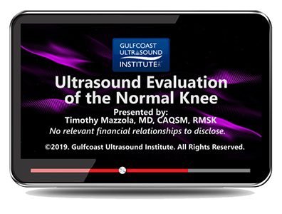
Gulfcoast: Ultrasound Evaluation of the Normal Knee


$15.00
VIDEO COURSE · Orthopedics · 2021
A focused ultrasound evaluation normal knee training video from Gulfcoast Ultrasound Institute — reviewing normal knee anatomy, ultrasound characteristics, scan techniques for the anterior, medial, lateral, and posterior compartments, and a complete routine examination protocol.
-
6topics
-
2021year
- Normal knee anatomy and its ultrasound characteristics
- Scan techniques for anterior, medial, lateral, and posterior compartments
- Complete routine knee examination protocol
- Limitations and pitfalls of knee ultrasound evaluation
Ultrasound evaluation normal knee — scan techniques and protocol from Gulfcoast
The ultrasound evaluation normal knee from Gulfcoast Ultrasound Institute provides a focused review of normal knee anatomy and its ultrasound characteristics. The training video walks through scan techniques for evaluating the anterior, medial, lateral, and posterior knee, demonstrates a complete routine examination protocol, and addresses the limitations and pitfalls that affect diagnostic accuracy. This is a practical, technique-driven resource for clinicians incorporating MSK ultrasound into their practice.
-
6topics
-
2021year
Inside this resource
Normal knee anatomy on ultrasound
The video opens with a systematic review of normal knee anatomy — bony landmarks, ligaments, tendons, cartilage, and fluid spaces — and how each structure appears on ultrasound. Understanding the normal sonographic appearance is the foundation for recognizing pathology, and this session provides the detailed baseline that sonographers need before tackling abnormal findings.
Scan techniques across all four compartments
The core of the training covers compartment-specific scan techniques. Anterior knee evaluation addresses the quadriceps tendon, patellar tendon, prepatellar bursa, and suprapatellar recess. Medial and lateral compartments examine collateral ligaments and meniscal margins. Posterior knee techniques assess the popliteal fossa, gastrocnemius-semimembranosus bursa, and Baker’s cyst evaluation. Each compartment is demonstrated with probe placement, orientation, and optimization tips.
Examination protocol and clinical application
A complete routine examination protocol ties the compartment-specific techniques into a reproducible workflow. The video addresses the limitations of knee ultrasound — what it cannot show compared to MRI — and common pitfalls such as anisotropy, acoustic shadowing, and misidentification of normal variants. Learning objectives include increasing confidence in performing and interpreting MSK ultrasound of the knee and applying standardized protocols to improve diagnostic and treatment accuracy.
Who this serves
- Point-of-care ultrasound users — structured technique training for knee evaluation in primary care, sports medicine, and rheumatology
- Diagnostic medical sonographers — focused knee protocol review for MSK ultrasound departments
- Emergency medicine and sports medicine physicians — practical scan techniques for bedside knee assessment
Before you buy
Is this about normal anatomy only?
Yes — this training video focuses on normal knee anatomy and scan techniques. It does not cover pathological findings such as meniscal tears or ligament injuries.
Who produces this content?
Gulfcoast Ultrasound Institute, a recognized provider of ultrasound education and training.
The chart
| Format | Video training |
|---|---|
| Publisher | Gulfcoast Ultrasound Institute |
| Department | Orthopedics, Radiology |
| Delivery | Instant download after checkout · lifetime re-access |
Verified original fileInstant downloadEncrypted checkout
5 reviews for Gulfcoast: Ultrasound Evaluation of the Normal Knee
Add a review
Related products



Orthopedics

Kevin M. –
Concise and practical. The compartment-by-compartment scan technique walkthrough gave me a reproducible workflow for bedside knee ultrasound. The pitfalls section saved me from anisotropy errors.
Monica R. –
Solid technique video from Gulfcoast. The probe placement tips for the medial and lateral compartments were particularly helpful. Good value for a focused MSK ultrasound resource.
Laura B. –
Good foundation in normal knee ultrasound anatomy. The anterior and posterior knee scanning demonstrations were clear. I would have liked some abnormal examples but as a normal-anatomy review it does its job well.
Samir H. –
Exactly what I needed to build confidence in MSK ultrasound. The protocol demonstration at the end ties everything together. Short, focused, and clinically useful.
Alan P. –
Well-produced but basic. If you already have some MSK ultrasound experience, this may feel introductory. For beginners it is a solid starting point for knee sonography.