


DentistryOnline courseDigital Dentistry
CBCT and dental radiology from first scan to full-practice integration — 80 hours from more than 30 imaging experts
Buying a cone beam unit is the easy part; reading every slice with confidence takes longer. DentalXP Dental Radiology and CBCT gathers lectures by 31 clinicians, radiologists and imaging specialists — among them Michael Bornstein, Martin D. Levin, Randolph R. Resnik, Bruno Azevedo and Ingrid Różyło-Kalinowska — into five modules and about 80 hours of video. The range runs from panoramic basics and normal anatomy to implant planning, endodontic diagnosis, TMJ imaging and merging volumes with intraoral scans.
- 5modules
- 81lectures listed
- 80 h 34 minof video
- 31speakers
What you’ll be able to do
- Choose the right field of view, protocol and machine for each diagnostic question
- Read mandibular and maxillary CBCT anatomy and spot variants before they cause complications
- Recognise and document incidental findings on panoramic and cone beam images
- Plan implants, sinus lifts and guided surgery directly from the volume
- Apply 3D imaging to endodontic diagnosis and TMJ assessment, including when MRI is the better choice
- Combine CBCT with intraoral scans for a digital treatment plan
Inside the course
Foundations and equipment choices
Around 17 hours on how cone beam imaging works, its strengths and limits, capture protocols, panoramic and extraoral technique, and how to pick a CBCT or OPG unit for an implant practice.
Interpretation and anatomy
Regional anatomical reviews of both jaws, crash courses for new users, panoramic interpretation, common oral pathology on 3D scans and a guided read-through of a real volume.
Implantology, surgery and endodontics
The largest clinical block covers implant planning tools, full-arch and guided protocols, the esthetic zone and sinus grafting. A shorter module turns to periapical diagnosis, outcome assessment, TMJ interpretation and an MRI primer.
Running 3D imaging in a real practice
Almost 25 hours on patient selection for general dentists, interdisciplinary planning, in-office guide fabrication, CAD/CAM links, paediatric imaging, full-mouth reconstruction and long-term follow-up of cases.
Built for
- General dentists buying or starting with CBCTPractical guidance on selection, protocols and responsible reading.
- Implant surgeons and periodontistsMany hours of volume-based planning for grafting, guides and full arches.
- Endodontists and TMD cliniciansDedicated lectures on 3D diagnosis of roots, lesions and joints.
Curriculum
Module 1: Foundations of 3D Imaging & CBCT (17 h 31 min) 20 lessons
- 3(D)entistry Roadmap Paradigm Shift Ahead
- 3D Dentistry Roadmap Paradigm Shift Ahead
- Basic understanding of cone beam CT
- Fundamentals of CBCT Imaging; Anatomy and Anatomical Variations
- Cone Beam Computed Tomography (CBCT) as a diagnostic aid in dentistry
- Cone Beam Computed Tomography (Vol. 1, Issue 1)
- CBCT Advantages and Limitations
- Benefits and limitations of Cone-Beam Computed Tomography in Clinical Practice
- CBCT Protocols
- CBCT selection
- CBCT Basics
- CBCT Course Intro
- CBCT Is Now The Right Time For Your Practice
- Panoramic Imaging
- Panoramic Radiographs in dental practice
- Panoramic Radiography and Extraoral Radiography
- Dental Radiology: Technique
- Essentials of CBCT: What Images to Capture and When
- How to select the best CBCT & OPG Machines for your Implant Practice
- Digital Dental Radiology – uncovering the mystery
Module 2: CBCT Anatomy, Interpretation & Incidental Findings (13 h 17 min) 15 lessons
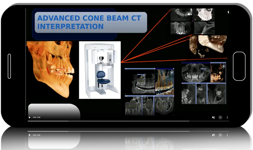
- Advanced CBCT Interpretation
- CBCT Anatomical Review of the Mandible
- CBCT Anatomical Review of the Maxillary Region
- CBCT Anatomy and Incidental Findings
- CBCT Interpretation Crash Course for the New User
- An Hour with an Expert -Understanding Radiology in CBCT
- Back to Basics Panoramic Radiographic Interpretation
- Bornstein Radiological analysis
- CBCT in Action: Common Oral Pathologies
- CBCT Stranger to Best Friend
- Cone Beam CT: Incidental Findings
- Identifying CBCT Mandibular Anatomic Variants To Prevent Complications
- Incidental findings in Pano
- Open your eyes to 3d introduction to cbct interpretation
- Example CBCT scan being interpreted
Module 3: CBCT Applications in Oral Implantology & Surgery (16 h 03 min) 16 lessons
- 3D Imaging and its Applications in Oral Implantology
- 7 Ways to Improve Patient Care with 3D Implant Dentistry and Guided Surgery
- CBCT and Implantology
- CBCT Guided Full Arch Dental Implant
- CBCT IMAGING IN GUIDED SURGERY
- CBCT Implant Treatment Planning by Mr. Christian Hall
- CBCT in Dental Implantology : World Implant Expo 2021
- Cone Beam CT – basic implant planning tools and strategy
- Cone Beam CT Interpretation for Zest LODI Implants by Robert Chen
- Dental Treatment: CBCT Imaging in Implant Dentistry
- Dental Webinar Series – Radiology for Oral Implantology by
- Diagnostic Imaging for Implant Treatment Planning
- Planning Implant Cases with CBCT
- Applications of CBCT for Dental Implants
- CBCT In Action: Sinus Lift Surgical Planning
- CBCT In Action: Implants in the Esthetic Zone
Module 4: CBCT Applications in Endodontics & Other Dental Specialties (8 h 55 min) 9 lessons
- CBCT and endo
- CBCT IN ENDO Carestream 4.8
- CBCT in Endodontics – A clinical approach with scientific evidence
- CBCT Interpretation in Endodontics II
- Importance of CBCT in clinical endodontics
- Diagnosis and Outcome in Endodontics in the 3D Imaging Era
- CBCT TMJ Interpretation
- MRI of TMJ
- MRIs made easy
Module 5: Integrating 3D Imaging into Practice Workflow (24 h 48 min) 21 lessons
- 3D Imaging Complete Vision for Every Practice
- 3D Imaging Known Knowns, Known Unknowns and Unknown Unknowns
- A Completely Digital Workflow for in-office fabrication of surgical
- Advanced Treatment Planning With CBCT
- CBCT CadCam Restorative Technologies in Clinical Dentistry
- CBCT as an Interdisciplinary Diagnostic and Treatment Planning
- CBCT For General Practitioners
- CBCT in Action: Patient Selection in the General Practice
- Launching Cone Beam CT Technology in Your Dental Office Pt 1
- Maximizing CBCT Technology in Today’s Practice
- Merge CBCT with Intraoral Scans to Achieve High-Definition Digital Diagnosis & Treatment Planning
- Using digital imaging for tim
- On CBCT and or Digital Dentistry in OMF
- Diagnostic Oral Radiographic Images in the Pediatric Patient- From A to Z
- CBCT and full mouth reconstruction
- CBCT Planning and 5 year follow up of cases done using Basal and Compressive Implants (ODC Dubai 20)
- CBCT report: healing
- CBCT Treatment Planning
- An Hour with an Expert- Panoramic Techniques and Common Errors
- CBCT Party at Home
- Dual CT scan
Before you buy
Is it one continuous course or a collection of lectures?
It is a curated compilation of talks and webinars from many speakers, grouped by theme, so style, recording quality and some content overlap vary between lectures.
How many lectures are actually included?
The headline figure is 117 lessons, but the published curriculum names 81 titles; the full list is shown above.
Do I need my own CBCT unit?
No. The interpretation and planning lectures are useful whether you own a scanner or refer patients to an imaging centre.
Do I get CME credit or a certificate?
No. Clinicapedia provides the educational content only — no CME or CE credits, MOC points or certificates of completion are included with this product.
Specifications
| Format | Online video course (lecture collection) |
|---|---|
| Instructors | 31 speakers, incl. Bornstein, Levin, Resnik, Azevedo |
| Contents | 5 modules · 81 listed lectures |
| Duration | 80 h 34 min |
| Access | Lifetime, self-paced |
| Department | Dentistry |
| Delivery | Instant download · lifetime re-download |
Related products




Dentistry

Dentistry

Anesthesiology

