


DentistryOnline courseGnathology
Read TMJ and maxillofacial images with confidence — MRI, CBCT, ultrasound, panoramic and intraoral radiography with Kaan Orhan
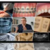
Taught by Ankara University radiology professor Kaan Orhan, Diagnostic Radiology: Focus on TMJ & Maxillofacial is a 10-lesson, 13.5-hour block that opens with the temporomandibular joint and then widens to general dentomaxillofacial imaging. You study joint anatomy and pathology on every major modality, then learn how each technique works, when to request it, how to acquire a good image and what the common findings look like.
- 10lessons
- 13 h 30 minof video
- 5imaging modalities
What you’ll be able to do
- Identify disc displacement with and without reduction, including partial, lateral and posterior patterns
- Recognise effusion, marrow oedema, erosions and osteoarthritic change in the condyle
- Choose between MRI, CBCT, ultrasound, OPG and intraoral films for a given clinical question
- Take better periapical, bitewing and occlusal radiographs with paralleling, bisecting and SLOB techniques
- Spot and avoid common panoramic and CBCT artifacts
- Screen for cysts, tumours, fibro-osseous lesions and osteonecrosis on a panoramic view
Inside the course
The joint first
Five lessons cover TMJ anatomy, histology and biomechanics together with the clinical muscle examination, then the full spectrum of internal derangement, degenerative, developmental and traumatic conditions, each illustrated with cases on CT, CBCT, MRI and ultrasound.
Everyday radiography done properly
A two-hour lesson on intraoral imaging explains projection geometry, sharpness, distortion, receptor positioning and paediatric exposures, followed by a practical session on normal and pathological radioanatomy of teeth and the periapical region.
Panoramic, CBCT, MRI and ultrasound fundamentals
The final four lessons treat each modality in turn: patient positioning and focal trough for OPG, detector design and artifacts in CBCT, MRI safety and parameter choice, and probe technique for ultrasound of the maxillofacial soft tissues.
About Kaan Orhan
Dean of dentistry at Ankara University, he has over 500 indexed publications and has led both the European and the International academies of dentomaxillofacial radiology.
Built for
- TMD and orofacial pain cliniciansImaging criteria that confirm or rule out joint pathology.
- Orthodontists and surgeonsKnow which scan to order and what the report should show.
- General dentistsA refresher on intraoral and panoramic technique plus the lesions that must not be missed.
Curriculum
Lesson 1: Structural anatomy, microscopic features, functional physiology and mechanical behavior of the temporomandibular joint (51 min) 8 topics
- Imaging-based overview of the temporomandibular joint (TMJ) and adjacent regions
- Review of typical TMJ morphology: bony landmarks and principal soft tissue elements
- Detailed look at the synovial lining, joint capsule, and stabilizing ligaments
- Structure and position of the articular disc in both sagittal and coronal planes
- Characteristics and function of the retrodiscal tissue
- Blood supply and related vascular structures of the TMJ
- Functional mechanics of the joint during movement
- Practical clinical assessment of the patient: muscle examination, palpation techniques, and identification of tender areas
Lesson 2: Disorders of the TMJ: disc position changes, joint degeneration and subluxation (1 h 16 min) 13 topics
- Diagnostic criteria for classifying TMJ disc (ADD) position and the different patterns of disc displacement
- Disc displacement with reduction and disc displacement without reduction in the TMJ
- Incomplete or segmental displacement of the articular disc
- Lateral (sideways) displacement patterns of the TMJ disc
- Posteriorly positioned disc displacement
- Identification and interpretation of joint effusion
- Bone marrow edema and its relationship to osteoarthrosis
- Degenerative joint changes characteristic of osteoarthritis in the TMJ
- Developmental abnormalities of the TMJ, including hypoplasia and bifid or trifid condyles
- Traumatic injuries of the TMJ, including joint luxations
- Overview of temporomandibular joint disorders
- Recognition of erosive changes and cystic lesions
- Discussion and analysis of selected clinical cases
Lesson 3: TMJ Imaging Essentials: From Traditional Methods to CT and CBCT in Joint Diagnosis (1 h) 8 topics
- Traditional methods used in dental and maxillofacial radiology
- Fundamentals and clinical use of panoramic X-ray imaging
- Tomographic imaging approaches, including conventional tomography, computed tomography (CT), and cone beam computed tomography (CBCT)
- How CBCT systems operate: image acquisition workflow, technical functioning, and radiation dose considerations
- Key technical characteristics and performance parameters of CBCT units
- Recognition and interpretation of common CBCT image artifacts
- CBCT-based anatomical assessment of the maxillofacial region, linked to corresponding gross anatomical structures
- Discussion and interpretation of selected clinical cases
Lesson 4: Temporomandibular Joint Imaging with MRI (53 min) 8 topics
- Historical development and key milestones in MRI imaging
- Core principles and essential concepts of MRI diagnostics
- How the MRI scanner works and step-by-step overview of the examination process
- Foundational anatomy of the TMJ as seen on MRI
- Detailed MRI anatomy of the TMJ disc and associated structures
- Typical imaging appearances of common TMJ pathologies
- MRI assessment of the masticatory muscles
- Relationship of the TMJ with the temporal bone and adjacent soft tissues on MRI
Lesson 5: USG of TMJ (1 h 10 min) 12 topics
- Orientation to maxillofacial ultrasound anatomy: key areas and landmark structures
- Survey of the major muscles and salivary glands in the region
- Visualization of vascular anatomy using static and dynamic MRI sequences
- Practical ultrasound techniques linked to gross anatomical relationships
- Assessment of the floor of the mouth in both transverse and longitudinal planes
- Normal appearance of the salivary glands on MRI in transverse and longitudinal views
- Typical MRI images of normal and reactive lymph nodes
- MRI and ultrasound of the TMJ: anatomical relationships and joint components
- When and why to request an ultrasound study
- Comparing normal and reactive findings on ultrasound with CBCT
- Identification of TMJ disorders on ultrasound and comparison with MRI and CBCT findings
- Discussion and analysis of a representative clinical case
Lesson 6: Intraoral X‑ray methods, when to use them, image acquisition, and radiographic anatomy (2 h 10 min) 15 topics
- Fundamentals of intraoral radiography and essential image quality standards
- Core concepts of projection geometry as they apply to radiographic imaging
- Factors influencing image sharpness and resolution, and strategies to enhance them
- Causes and recognition of image magnification and distortion in size and shape
- Periapical imaging using the paralleling technique
- A complete, stepwise exposure protocol from preparation to image capture
- Correct positioning of the receptor for various intraoral projections
- Indications, benefits, and limitations of the bisecting‑angle technique
- Application of the Donovan technique
- Application of the Le Master technique
- Application of the SLOB (Same Lingual, Opposite Buccal) technique
- Use of vertical and horizontal bitewing receptors and when each is preferred
- Occlusal radiographs and their clinical indications
- Intraoral radiographic examination in pediatric patients, including mobile radiography
- Specific clinical and technical considerations in special situations
Lesson 7: Getting Started with OPG: how to take the image, when to request it, step-by-step scanning, key anatomical landmarks, and typical pathological findings (1 h 24 min) 34 topics
- Overview of orthopantomography: what it is, how it works, and how it evolved over time in dental imaging
- Concept of the tomographic focal layer in panoramic radiography
- Digital detector technologies used in panoramic units and their main characteristics
- When panoramic radiography is recommended in clinical practice
- Benefits and limitations of panoramic examinations compared with other imaging methods
- Step‑by‑step panoramic workflow: correct patient alignment, tongue placement, and frequent technical errors
- Role and impact of cervical spine extension on image quality
- Typical image artifacts and how to recognize them:
- Ghost formations
- Superimposed foreign objects
- Distortions related to metallic materials
- Artifacts due to the patient closing incorrectly
- Blurring caused by movement
- Effects of the lead apron
- Errors linked to improper tongue position
- Systematic identification of anatomical landmarks: comprehensive review of bony and soft tissue structures on the OPG
- Clinical reasons for selecting panoramic imaging
- Diagnostic questions that demand a wide overview of both jaws
- Interpretation of panoramic views in various clinical scenarios, including:
- Assessment of facial and jaw trauma
- Localization of third molars
- Investigation of widespread dental or bone pathology
- Evaluation of suspected or confirmed large jaw lesions
- Analysis of tooth formation and eruption
- Detection of retained teeth or residual roots in edentulous areas
- Hard-tissue–related temporomandibular joint conditions (condylar region)
- Developmental disturbances of the jaws and dentition
- Recognition of disease on panoramic radiographs:
- Cystic jaw lesions
- Benign neoplastic processes
- Malignant tumors
- Fibro‑osseous conditions
- Jaw osteonecrosis, including osteomyelitis, MRONJ, and ORN
- Discussion and interpretation of a variety of real clinical panoramic cases
Lesson 8: CBCT Basics: How to Scan, When to Use It, Key Anatomy, and Typical Pathologies (1 h 26 min) 5 topics
- Fundamental concepts behind CBCT imaging
- Frequently encountered pathological findings in CBCT scans
- How dentomaxillofacial CBCT machines work, with a focus on how their various x‑ray detection designs differ
- Types of image artifacts and how they appear
- Key distinctions between panoramic radiography (OPG) and CBCT imaging
Lesson 9: MRI basics: imaging methods, when to order a scan, examination workflow, key anatomy, and frequently encountered diseases (1 h 45 min) 5 topics
- Core principles and fundamentals of MRI
- Using anatomical and physiological understanding to guide MRI patient positioning
- Recognition of frequently encountered disease patterns in MRI studies
- Identification and understanding of image artifacts in MRI
- Selection of scan parameters for superior image quality, plus practical use of ferromagnetic safety and contrast media to safeguard patients, technologists, and all personnel in the MRI environment
Lesson 10: Fundamentals of ultrasound imaging and radiography: techniques, clinical indications, scanning approach, normal anatomy, and key pathological findings (1 h 24 min) 3 topics
- Fundamental principles of ultrasound imaging
- Probe placement and scanning techniques for the DMFR region and adjacent soft tissues
- Identification and interpretation of pathological findings on ultrasound
Before you buy
Is it only about the TMJ?
No. The first five lessons centre on the joint; the remaining five are general introductions to intraoral, panoramic, CBCT, MRI and ultrasound imaging.
Is the hands-on part practical?
The radioanatomy session in lesson 6 is an interpretation exercise on recorded image examples rather than physical training.
Do I get CME credit or a certificate?
No. Clinicapedia provides the educational content only — no CME or CE credits, MOC points or certificates of completion are included with this product.
Specifications
| Format | Online video course |
|---|---|
| Instructor | Kaan Orhan |
| Contents | 10 lessons |
| Duration | 13 h 30 min |
| Access | Lifetime, self-paced |
| Department | Dentistry |
| Delivery | Instant download · lifetime re-download |
Related products



Anesthesiology




Dentistry
