
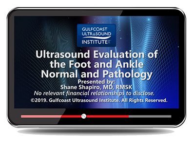
Gulfcoast: Ultrasound Evaluation of the Foot and Ankle: Normal and Pathology
$15.00
VIDEO COURSE · Gulfcoast Ultrasound Institute
This foot ankle ultrasound evaluation training covers anatomy, scan techniques, normal ultrasound characteristics, and commonly seen pathology — from Achilles tendinopathy and plantar fasciitis through tarsal tunnel syndrome and Morton neuroma, with diagnostic criteria for each condition.
-
20pathology topics
- Normal anatomy of anterior, medial, lateral, and posterior ankle
- Patient positioning and scan technique for systematic foot-ankle evaluation
- Tendinopathy, ligament tears, and entrapment neuropathy assessment
- Diagnostic criteria for 20 commonly seen foot and ankle conditions
Foot and ankle ultrasound evaluation for normal anatomy and pathology
This foot ankle ultrasound evaluation training from Gulfcoast provides a systematic review of foot and ankle anatomy, scanning technique, and commonly seen pathology. The course establishes the normal ultrasound characteristics across anterior, medial, lateral, and posterior approaches before addressing the conditions — tendinopathy, ligament injury, entrapment neuropathy, and degenerative disease — that clinicians encounter most frequently in foot and ankle practice.
Inside this resource
Normal anatomy and patient positioning
The course opens with normal foot and ankle anatomy across four scanning approaches — anterior, medial-lateral, and posterior-plantar. Patient positioning and scan technique are demonstrated for each window, establishing the systematic approach that ensures complete evaluation. This normal anatomy baseline serves as the reference standard for recognising pathological findings.
Tendon pathology: Achilles, peroneal, and flexor assessment
The tendon modules cover Achilles tendinopathy and rupture evaluation, peroneal tendinopathy with subluxation and dislocation assessment, peroneus quartus identification, and flexor hallucis longus tendinitis. Each condition is presented with its ultrasound characteristics and the diagnostic criteria that distinguish normal variation from pathology.
Ligament, nerve, and soft tissue pathology
Ligament assessment covers ATFL tears and spring ligament evaluation. Nerve pathology addresses tarsal tunnel syndrome assessment. Soft tissue conditions include retrocalcaneal bursitis, plantar fasciitis, Morton neuroma, and midfoot degenerative joint disease. Hallux rigidus evaluation completes the pathology coverage.
Effusion detection and diagnostic criteria
Joint effusion assessment provides the scanning technique and criteria for detecting fluid collections in the foot and ankle — a finding that may indicate inflammatory, traumatic, or degenerative joint disease. The diagnostic criteria presented throughout the course build a systematic framework for clinical decision-making.
Who this serves
- Sonographers in foot and ankle practice — develop systematic evaluation skills for the full range of foot and ankle pathology
- Podiatrists and orthopaedic surgeons — refine ultrasound interpretation for diagnostic and procedural applications
- Sports medicine clinicians — build pattern recognition for athletic foot and ankle injury assessment
Before you buy
Does this cover both normal anatomy and pathology?
Yes — the course begins with normal anatomy and scan technique before addressing 20 commonly seen foot and ankle pathologies with diagnostic criteria.
Which pathologies are covered?
Achilles and peroneal tendinopathy, ATFL tears, tarsal tunnel syndrome, plantar fasciitis, Morton neuroma, retrocalcaneal bursitis, and midfoot degenerative disease among others.
The chart
| Format | Video lecture (MP4) |
|---|---|
| Publisher | Gulfcoast Ultrasound Institute |
| Department | Gulfcoast Ultrasound Institute |
| Delivery | Instant download after checkout · lifetime re-access |
Verified original fileInstant downloadEncrypted checkout
6 reviews for Gulfcoast: Ultrasound Evaluation of the Foot and Ankle: Normal and Pathology
Add a review
Related products
Gulfcoast Ultrasound Institute
AIUM Prenatal Ultrasound Abnormalities of the Chest, Abdomen, and Pelvis With Postnatal Correlation
Gulfcoast Ultrasound Institute
Gulfcoast Ultrasound Institute
Gulfcoast Ultrasound Institute
Gulfcoast Ultrasound Institute
Gulfcoast Ultrasound Institute
Gulfcoast Dynamic Musculoskeletal Ultrasound Imaging (Videos+PDFs)
Gulfcoast Ultrasound Institute
GCUS Ultrasound Evaluation of the Fetal Heart: Basic and Advanced Techniques 2021 (VIDEOS)

Gulfcoast Ultrasound Institute
Carlos M. –
Downloaded for a foot and ankle scanning refresher. The normal anatomy baseline before pathology assessment is a well-organised approach.
Marcus J. –
Twenty pathology topics in a single comprehensive training. The Achilles and peroneal tendon sections are particularly well structured with clear diagnostic criteria.
Hannah M. –
Practical for foot and ankle scanning. The tarsal tunnel syndrome and plantar fasciitis modules address conditions I see regularly in clinic.
drtomh –
Good balance of normal anatomy and pathology. The patient positioning demonstrations for each scanning approach are immediately practical.
Lynn C. –
Solid content but covering 20 pathologies in a single lecture means some topics receive limited time. The ligament section could use more depth.
Fatima R. –
Excellent systematic coverage of foot and ankle ultrasound. The Morton neuroma and midfoot DJD sections are particularly well presented.