
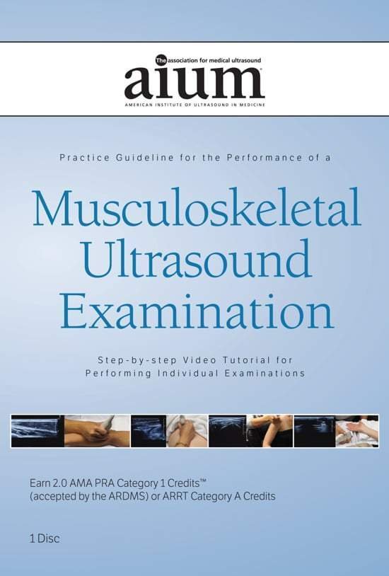
AIUM PRACTICE PARAMETER FOR Musculoskeletal Ultrasound Examination- Step-by-Step Video Tutorial


$15.00
VIDEO COURSE · AIUM · MSK Guidelines
This MSK ultrasound examination technique tutorial uses live models to demonstrate the sonographic evaluation and standardised views for each joint as recommended by the AIUM practice guideline for musculoskeletal ultrasound — designed to illustrate the correct implementation of clinical practice guideline recommendations.
-
1video tutorial
- Joint-by-joint sonographic evaluation per AIUM guideline
- Live model demonstrations of standardised scanning views
- Correct implementation of clinical practice recommendations
- Focus on patient care, medical knowledge, and practice-based learning
MSK ultrasound examination technique per AIUM practice guideline
This MSK ultrasound examination technique tutorial from the AIUM demonstrates the sonographic evaluation and view acquisition for each joint as recommended by the AIUM practice guideline for musculoskeletal ultrasound. Using live models, the programme illustrates the correct implementation of clinical practice guideline recommendations — covering patient care, medical knowledge, and practice-based learning improvement.
Inside this resource
Joint-by-joint evaluation technique
The tutorial walks through the sonographic assessment of each major joint using the standardised views specified by the AIUM musculoskeletal practice guideline. Each joint is demonstrated with a live model — showing transducer placement, patient positioning, and the anatomical landmarks that confirm a complete and adequate examination.
Standardised view acquisition
For every joint, the tutorial demonstrates how to acquire the standardised views that form the basis of a complete musculoskeletal ultrasound examination. The emphasis is on reproducibility — the scanning sequence and patient positioning that allow consistent, reliable image acquisition across different body habitus types.
Practice-based learning and improvement
Beyond the technical scanning elements, the tutorial addresses the learner attributes that support ongoing competency development — patient care principles during MSK scanning, the medical knowledge foundation for image interpretation, and how systematic practice leads to improved scanning proficiency over time.
Who this serves
- Sonographers in MSK practice — align scanning technique with AIUM musculoskeletal guidelines
- Physicians learning MSK ultrasound — build foundational joint evaluation competency with live demonstrations
- Radiology trainees — understand the standardised approach recommended for musculoskeletal ultrasound examinations
Before you buy
Does this use live model demonstrations?
Yes — the tutorial uses live models to demonstrate transducer placement, patient positioning, and view acquisition for each joint.
Which joints are covered?
The tutorial demonstrates the standardised views for the major joints as specified by the AIUM practice guideline for musculoskeletal ultrasound examination.
The chart
| Format | Video tutorial (MP4) |
|---|---|
| Publisher | AIUM |
| Department | Gulfcoast Ultrasound Institute |
| Delivery | Instant download after checkout · lifetime re-access |
Verified original fileInstant downloadEncrypted checkout
5 reviews for AIUM PRACTICE PARAMETER FOR Musculoskeletal Ultrasound Examination- Step-by-Step Video Tutorial
Add a review
Related products
Gulfcoast Ultrasound Institute
Gulfcoast Advanced Ultrasound Techniques for the Foot and Ankle (Videos+PDFs)
Gulfcoast Ultrasound Institute

Gulfcoast Ultrasound Institute
AIUM Prenatal Ultrasound Abnormalities of the Chest, Abdomen, and Pelvis With Postnatal Correlation
Gulfcoast Ultrasound Institute

Gulfcoast Ultrasound Institute
Gulfcoast Ultrasound Institute
Gulfcoast Ultrasound Institute
AIUM VIDEO HD Musculoskeletal Ultrasound of Sports Injuries and Hernias

Gulfcoast Ultrasound Institute
Lynn C. –
Downloaded as a review before certification. The practice-based learning focus adds a valuable educational framework to the technical demonstrations.
M. Alvarez –
Live model demonstrations are the strength of this tutorial. Seeing transducer placement and patient positioning in real time is far more useful than static diagrams.
James R. –
Practical and well-structured. The standardised view acquisition method is presented clearly — helps establish consistent scanning habits early.
Priya –
Useful content but a single tutorial covers a lot of ground. Would have appreciated more time on the shoulder and hip joints specifically.
Marcus J. –
Good reference for aligning MSK scanning technique with AIUM guidelines. The joint-by-joint approach makes it easy to review specific regions.