
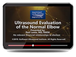
Gulfcoast: Ultrasound Evaluation of the Normal Elbow
$15.00
VIDEO COURSE · Gulfcoast Ultrasound Institute
This normal elbow ultrasound evaluation training provides an introduction to elbow anatomy, scanning technique, and the sonographic characteristics of normal structures — covering anterior, medial, and lateral compartments with practical scan demonstrations.
-
14topic areas
- Anterior elbow anatomy and scanning approach
- Medial elbow: common flexor tendon and ulnar nerve assessment
- Lateral elbow: radial collateral ligament and radial nerve evaluation
- Scan demonstration with practical protocol application
Normal elbow ultrasound evaluation for anatomy and scanning technique
This normal elbow ultrasound evaluation training from Gulfcoast introduces the anatomy, scanning protocols, and sonographic characteristics associated with the normal elbow. Fourteen topic areas cover anterior, medial, and lateral compartments — including the pronator window, distal biceps, ulnar nerve, and radial nerve — with practical scan demonstrations that establish the systematic approach for routine elbow assessment.
Inside this resource
Anterior elbow anatomy and scanning approach
The anterior module covers the biceps tendon, distal biceps insertion, and the pronator window — the key anatomical landmarks for anterior elbow assessment. Joint evaluation addresses the anterior recess and the scanning technique that reveals effusion and synovial thickening in the elbow joint.
Medial and lateral compartment assessment
Medial elbow evaluation covers the common flexor tendon and ulnar nerve assessment — the structures most relevant to medial elbow pain and cubital tunnel evaluation. Lateral elbow scanning addresses the radial collateral ligament, annular ligament, and radial nerve assessment — the structures involved in lateral epicondylitis and radial tunnel syndrome.
Posterior elbow and scan demonstration
The posterior module covers triceps tendon evaluation and the posterior elbow joint assessment. A practical scan demonstration integrates all compartments into a systematic protocol — showing transducer placement, anatomical landmark identification, and the systematic approach that ensures complete elbow evaluation.
Who this serves
- Sonographers learning elbow MSK scanning — build foundational anatomy knowledge and scanning protocol competency
- Physicians new to elbow ultrasound — establish systematic evaluation skills with normal anatomy as the baseline
- Radiology trainees — develop elbow ultrasound technique with structured protocol demonstration
Before you buy
Does this cover pathology or normal anatomy only?
Focused on normal anatomy and scanning technique — the baseline knowledge needed before identifying pathological findings in elbow ultrasound.
Which anatomical regions does this cover?
Anterior, medial, lateral, and posterior — including the pronator window, distal biceps, ulnar nerve, radial nerve, and scan demonstration.
The chart
| Format | Video lecture (MP4) |
|---|---|
| Publisher | Gulfcoast Ultrasound Institute |
| Department | Gulfcoast Ultrasound Institute |
| Delivery | Instant download after checkout · lifetime re-access |
Verified original fileInstant downloadEncrypted checkout
6 reviews for Gulfcoast: Ultrasound Evaluation of the Normal Elbow
Add a review
Related products
Gulfcoast Ultrasound Institute
Gulfcoast Ultrasound Institute
Gulfcoast Upper Extremity Musculoskeletal Ultrasound Protocols (Videos)
Gulfcoast Ultrasound Institute
AIUM VIDEO HD Musculoskeletal Ultrasound of Sports Injuries and Hernias

Gulfcoast Ultrasound Institute

Gulfcoast Ultrasound Institute
Gulfcoast Ultrasound Institute

Gulfcoast Ultrasound Institute
Gulfcoast Ultrasound Institute
Gulfcoast Advanced MSK Ultrasound-Guided Procedures for Rehabilitation Medicine
Natalie F. –
Downloaded as a foundation before tackling elbow pathology courses. The normal baseline approach is exactly what I needed for building scanning competency.
Priya –
Clear introduction to normal elbow anatomy with scanning demonstrations. The pronator window and ulnar nerve sections are particularly well presented.
Wei L. –
Practical for learning elbow ultrasound technique. The radial nerve and lateral collateral ligament assessment are clearly demonstrated.
Sarah K. –
Good foundational elbow scanning course. The systematic protocol approach works well for building consistent scanning habits from the start.
Marcus J. –
Useful normal anatomy reference but would have liked more time on the posterior compartment. The triceps assessment felt brief.
M. Alvarez –
Excellent systematic introduction to elbow scanning. The scan demonstration integrating all compartments is the strongest section — practical and memorable.