
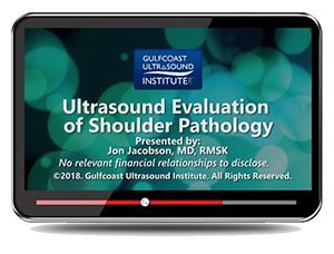
Gulfcoast: Ultrasound Evaluation of Shoulder Pathology
$15.00
VIDEO COURSE · Gulfcoast Ultrasound Institute
This shoulder pathology ultrasound evaluation training provides a comprehensive overview of commonly encountered shoulder pathology — covering rotator cuff tears from partial to massive, tendinosis, fatty infiltration, and the diagnostic criteria and scanning pitfalls that affect accurate shoulder assessment.
-
15topic areas
- Partial and full-thickness rotator cuff tear classification
- Tendinosis, fatty infiltration, and muscle atrophy assessment
- Joint and bursal effusion evaluation
- Scanning pitfalls and secondary findings in shoulder sonography
Shoulder pathology ultrasound evaluation for rotator cuff and tendon assessment
This shoulder pathology ultrasound evaluation training from Gulfcoast provides a comprehensive overview of the ultrasound characteristics and diagnostic criteria for commonly seen shoulder pathology. Fifteen topic areas cover the full spectrum of rotator cuff disease — from partial-thickness tears through massive cuff failure — along with tendinosis, fatty infiltration, muscle atrophy, and the secondary findings that guide clinical decision-making.
Inside this resource
Rotator cuff tear classification and evaluation
The course systematically addresses rotator cuff pathology — supraspinatus insertion tears, articular and bursal partial-thickness tears, large and massive full-thickness tears, and intrasubstance tears. Each tear pattern is presented with its ultrasound characteristics and the diagnostic criteria that distinguish it from adjacent pathology, building the pattern recognition needed for accurate classification.
Tendon quality assessment and secondary findings
Beyond tears, the training covers tendinosis evaluation, fatty infiltration staging, muscle atrophy assessment, and tendon volume loss — the chronic changes that influence surgical planning and prognosis. Joint and bursal effusion evaluation addresses the fluid collections that serve as secondary indicators of underlying cuff disease.
Diagnostic pitfalls and miscellaneous pathology
A dedicated section addresses the scanning pitfalls in rotator cuff sonography — the artefacts and anatomical variations that can mimic or mask pathology. Additional miscellaneous cuff pathology rounds out the diagnostic scope, covering conditions that fall outside the standard tear classification frameworks.
Who this serves
- Sonographers performing shoulder MSK scanning — develop systematic rotator cuff pathology recognition and classification skills
- Orthopaedic surgeons — refine ultrasound interpretation for surgical planning and cuff tear assessment
- Sports medicine physicians — build diagnostic accuracy for shoulder pathology in athletic populations
Before you buy
Does this cover normal shoulder anatomy?
Focused on pathology evaluation — rotator cuff tears, tendinosis, fatty infiltration, and secondary findings with diagnostic criteria.
Which rotator cuff tears are addressed?
Complete coverage: supraspinatus insertion tears, articular and bursal partial-thickness tears, large and massive full-thickness tears, and intrasubstance tears.
The chart
| Format | Video lecture (MP4) |
|---|---|
| Publisher | Gulfcoast Ultrasound Institute |
| Department | Gulfcoast Ultrasound Institute |
| Delivery | Instant download after checkout · lifetime re-access |
Verified original fileInstant downloadEncrypted checkout
6 reviews for Gulfcoast: Ultrasound Evaluation of Shoulder Pathology
Add a review
Related products
Gulfcoast Ultrasound Institute

Gulfcoast Ultrasound Institute
GCUS Introduction to Carotid Duplex/Color Flow Ultrasound 2022 (VIDEOS)

Gulfcoast Ultrasound Institute
GCUS Abdominal and Primary Care Ultrasound 2022 (Gulfcoast Ultrasound Institute) (Videos) 2022
Gulfcoast Ultrasound Institute
GCUS Introduction to Peripheral Vascular Ultrasound 2022 (VIDEOS)
Gulfcoast Ultrasound Institute
Gulfcoast Ultrasound Guided Prolotherapy: General Principles (Videos)

Gulfcoast Ultrasound Institute

Gulfcoast Ultrasound Institute
Gulfcoast Ultrasound Institute
Gulfcoast Upper Extremity Musculoskeletal Ultrasound Protocols (Videos)
Priya –
Downloaded for shoulder scanning competency. The systematic approach to cuff evaluation is immediately applicable in daily practice.
Sarah K. –
The rotator cuff tear classification section is thorough. Partial versus full-thickness tear distinction with ultrasound criteria is well demonstrated across all subtypes.
James R. –
Practical for shoulder MSK scanning practice. The pitfalls section helped me recognise errors I was making with artefact interpretation.
Wei L. –
Solid content but fifteen topics in a single lecture moves quickly. The secondary findings section felt rushed compared to the tear classification coverage.
M. Alvarez –
Good diagnostic criteria coverage. The fatty infiltration and muscle atrophy modules add clinical depth beyond just identifying tears.
Natalie F. –
Excellent resource for building shoulder pathology recognition. The intrasubstance tear evaluation is particularly well covered — a topic many courses skip.