
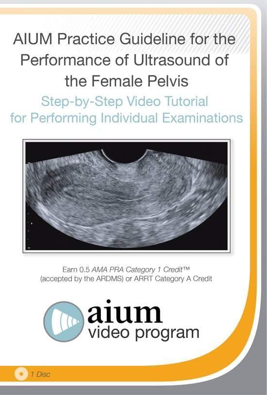
AIUM Practice Guideline for the Female Pelvis

$15.00
VIDEO COURSE · AIUM · Pelvic Imaging
This female pelvis ultrasound guideline tutorial covers the sonographic evaluation of the uterus, ovaries, cervix, and bladder — including the use of 3D ultrasound to visualise the coronal view of the uterus and pelvic floor structures as recommended by the AIUM practice guideline.
-
1video tutorial
- Complete ultrasound evaluation of the female pelvis
- 3D ultrasound for coronal uterine visualisation
- Pelvic floor assessment with sonographic technique
- Sonographic appearance of uterus, ovaries, cervix, and bladder
Female pelvis ultrasound guideline with 3D uterine and pelvic floor assessment
This female pelvis ultrasound guideline tutorial from the AIUM teaches the sonographic evaluation of the uterus, ovaries, cervix, and bladder — demonstrating a complete female pelvis ultrasound examination. The tutorial extends into 3D ultrasound applications for visualising the coronal view of the uterus and assessing pelvic floor anatomy, addressing the medical knowledge and patient care attributes needed for clinical competency.
Inside this resource
Sonographic appearance of female pelvic organs
The tutorial demonstrates the normal sonographic appearance of the uterus, ovaries, cervix, and bladder in both transabdominal and transvaginal approaches. Each organ is presented with the standardised views that allow systematic evaluation — endometrial assessment, ovarian follicle mapping, and cervical length measurement where clinically indicated.
3D ultrasound and coronal uterine visualisation
A dedicated section covers 3D ultrasound applications for the female pelvis — acquiring and interpreting the coronal view of the uterus, which is the standard plane for evaluating congenital uterine anomalies, endometrial cavity morphology, and IUD position. The technique for acquiring and rendering 3D volumes is demonstrated step by step.
Pelvic floor evaluation and complete examination protocol
The tutorial addresses pelvic floor assessment using ultrasound — evaluating levator ani integrity, bladder neck mobility, and the sonographic findings relevant to pelvic floor dysfunction. The complete examination protocol integrates all elements into a reproducible scanning sequence.
Who this serves
- Gynaecological sonographers — refine technique for complete female pelvis evaluation including 3D applications
- Gynaecologists — understand the standardised views and 3D acquisition for uterine anomaly assessment
- Trainees in obstetrics and gynaecology — build foundational pelvic ultrasound competency
Before you buy
Does this cover both 2D and 3D ultrasound?
Yes — the tutorial covers the standard 2D pelvic evaluation and extends into 3D applications for coronal uterine visualisation and pelvic floor assessment.
Is this a transabdominal or transvaginal approach?
The tutorial covers the complete examination, including both transabdominal and transvaginal technique as part of the systematic evaluation.
The chart
| Format | Video tutorial (MP4) |
|---|---|
| Publisher | AIUM |
| Department | Gulfcoast Ultrasound Institute |
| Delivery | Instant download after checkout · lifetime re-access |
Verified original fileInstant downloadEncrypted checkout
5 reviews for AIUM Practice Guideline for the Female Pelvis
Add a review
Related products
Gulfcoast Ultrasound Institute
Gulfcoast Ultrasound Institute
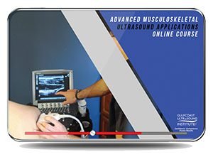
Advanced Musculoskeletal (MSK) Ultrasound Applications 2020 (VIDEOS)
Gulfcoast Ultrasound Institute
GCUS Introduction to Transcranial Doppler Ultrasound 2019 (VIDEOS)
Gulfcoast Ultrasound Institute
Gulfcoast Dynamic Musculoskeletal Ultrasound Imaging (Videos+PDFs)
Gulfcoast Ultrasound Institute
Gulfcoast Upper Extremity Musculoskeletal Ultrasound Protocols (Videos)

Gulfcoast Ultrasound Institute

Gulfcoast Ultrasound Institute
AIUM Prenatal Ultrasound Abnormalities of the Chest, Abdomen, and Pelvis With Postnatal Correlation

Gulfcoast Ultrasound Institute
Natalie F. –
Downloaded for a 3D ultrasound refresher. The coronal plane acquisition technique is presented in a way I can immediately apply in practice.
Priya –
The 3D coronal uterine view section alone justified the purchase. Step-by-step volume acquisition technique is clearly demonstrated.
Fatima R. –
Good coverage of both 2D and 3D pelvic assessment. The pelvic floor evaluation section adds depth beyond a basic gynaecology ultrasound tutorial.
Sarah K. –
Comprehensive female pelvis tutorial. The systematic approach to organ evaluation is well-structured — helpful for building a consistent scanning routine.
Carlos M. –
Content is solid but would have appreciated more case examples showing common uterine anomalies and their 3D appearance.